2AZQ

Crystal Structure of Catechol 1,2-Dioxygenase from Pseudomonas arvilla C-1


Experimental Data Snapshot

  • Method: X-RAY DIFFRACTION
  • Resolution: 2.65 Å
  • R-Value Free: 0.284 
  • R-Value Work: 0.229 
  • R-Value Observed: 0.238 

wwPDB Validation   3D Report Full Report


Ligand Structure Quality Assessment 


This is version 1.4 of the entry. See complete history


Literature

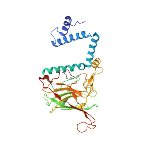
Structure of catechol 1,2-dioxygenase from Pseudomonas arvilla

Earhart, C.A.Vetting, M.W.Gosu, R.Michaud-Soret, I.Que, L.Ohlendorf, D.H.

(2005) Biochem Biophys Res Commun 338: 198-205

  • DOI: https://doi.org/10.1016/j.bbrc.2005.08.221
  • Primary Citation of Related Structures:  
    2AZQ

  • PubMed Abstract: 

    Catechol 1,2-dioxygenase was first studied by Hayaishi and colleagues in 1950. In 1967, catechol 1,2-dioxygenase from Pseudomonas arvilla C-1 (PaCTD) was chosen as a model system for the catecholic intradiol dioxygenases due to its activity, stability and expression level. Here we report the 2.65 A structure of the betabeta isozyme of PaCTD. The structure supports the hypothesis first made by Vetting and Ohlendorf [The 1.8A crystal structure of catechol 1,2-dioxygenase reveals a novel hydrophobic helical zipper as a subunit linker, Struct. Fold. Des. 8 (2000) 429-440.] that the catechol 1,2-dioxygenases are lipid binding proteins. The 5 amino-terminal helices involved in dimerization and forming the lipid binding site are shown to be plastic in their positions and orientations. The sequence differences between the alpha and beta polypeptides are located at the part of the monomers distant from dimerization surface and thus permit the formation of the 3 isozymes (alphaalpha, alphabeta, and betabeta) of PaCTD. The reported inactivation by sulfhydryl-modifying reagents is explained by the structure. The 10-residue Helix F (residues 203-212) is proposed to be central in communicating between the lipid binding site and the active site.


  • Organizational Affiliation

    Department of Biochemistry, Molecular Biology, and Biophysics, University of Minnesota, 6-155 Jackson Hall, 321 Church St. SE, Minneapolis, MN 55455, USA.


Macromolecules
Find similar proteins by:  (by identity cutoff)  |  3D Structure
Entity ID: 1
MoleculeChains Sequence LengthOrganismDetailsImage
catechol 1,2-dioxygenase311Pseudomonas putidaMutation(s): 0 
EC: 1.13.11.1
UniProt
Find proteins for Q51433 (Pseudomonas putida)
Explore Q51433 
Go to UniProtKB:  Q51433
Entity Groups  
Sequence Clusters30% Identity50% Identity70% Identity90% Identity95% Identity100% Identity
UniProt GroupQ51433
Sequence Annotations
Expand
  • Reference Sequence
Small Molecules
Ligands 2 Unique
IDChains Name / Formula / InChI Key2D Diagram3D Interactions
PCF
Query on PCF

Download Ideal Coordinates CCD File 
C [auth A]1,2-DIACYL-SN-GLYCERO-3-PHOSHOCHOLINE
C40 H80 N O8 P
KILNVBDSWZSGLL-KXQOOQHDSA-N
FE
Query on FE

Download Ideal Coordinates CCD File 
B [auth A]FE (III) ION
Fe
VTLYFUHAOXGGBS-UHFFFAOYSA-N
Experimental Data & Validation

Experimental Data

  • Method: X-RAY DIFFRACTION
  • Resolution: 2.65 Å
  • R-Value Free: 0.284 
  • R-Value Work: 0.229 
  • R-Value Observed: 0.238 
  • Space Group: C 2 2 21
Unit Cell:
Length ( Å )Angle ( ˚ )
a = 62.73α = 90
b = 71.52β = 90
c = 187.09γ = 90
Software Package:
Software NamePurpose
X-GENdata scaling
X-GENdata reduction
AMoREphasing
CNSrefinement

Structure Validation

View Full Validation Report


Ligand Structure Quality Assessment 


Entry History 

Deposition Data

Revision History  (Full details and data files)

  • Version 1.0: 2005-10-04
    Type: Initial release
  • Version 1.1: 2008-05-01
    Changes: Version format compliance
  • Version 1.2: 2011-07-13
    Changes: Derived calculations, Version format compliance
  • Version 1.3: 2017-10-11
    Changes: Refinement description
  • Version 1.4: 2023-08-23
    Changes: Data collection, Database references, Derived calculations, Refinement description