2A0I

F Factor TraI Relaxase Domain bound to F oriT Single-stranded DNA


Experimental Data Snapshot

  • Method: X-RAY DIFFRACTION
  • Resolution: 2.72 Å
  • R-Value Free: 0.272 
  • R-Value Work: 0.215 
  • R-Value Observed: 0.221 

wwPDB Validation   3D Report Full Report


This is version 1.6 of the entry. See complete history


Literature

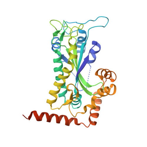
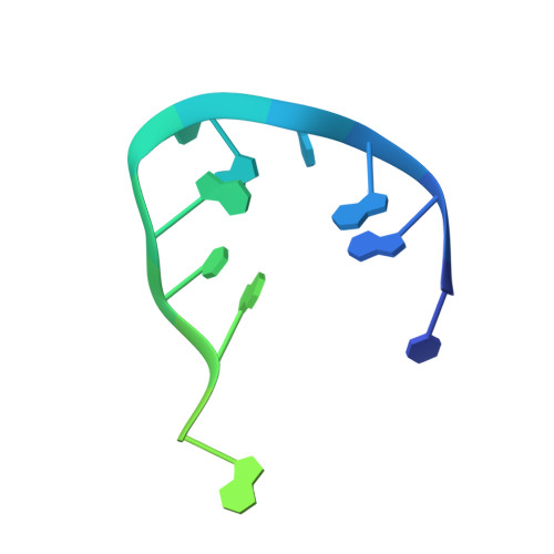
Inter- and intramolecular determinants of the specificity of single-stranded DNA binding and cleavage by the f factor relaxase.

Larkin, C.Datta, S.Harley, M.J.Anderson, B.J.Ebie, A.Hargreaves, V.Schildbach, J.F.

(2005) Structure 13: 1533-1544

  • DOI: https://doi.org/10.1016/j.str.2005.06.013
  • Primary Citation of Related Structures:  
    2A0I

  • PubMed Abstract: 

    The TraI protein of conjugative plasmid F factor binds and cleaves a single-stranded region of the plasmid prior to transfer to a recipient. TraI36, an N-terminal TraI fragment, binds ssDNA with a subnanomolar K(D) and remarkable sequence specificity. The structure of the TraI36 Y16F variant bound to ssDNA reveals specificity determinants, including a ssDNA intramolecular 3 base interaction and two pockets within the protein's binding cleft that accommodate bases in a knob-into-hole fashion. Mutagenesis results underscore the intricate design of the binding site, with the greatest effects resulting from substitutions for residues that both contact ssDNA and stabilize protein structure. The active site architecture suggests that the bound divalent cation, which is essential for catalysis, both positions the DNA by liganding two oxygens of the scissile phosphate and increases the partial positive charge on the phosphorus to enhance nucleophilic attack.


  • Organizational Affiliation

    Department of Biology, The Johns Hopkins University, 3400 North Charles Street, Baltimore, Maryland 21218, USA.


Macromolecules

Find similar proteins by:  (by identity cutoff)  |  3D Structure
Entity ID: 2
MoleculeChains Sequence LengthOrganismDetailsImage
TraI proteinB [auth A]330Escherichia coliMutation(s): 1 
Gene Names: traI
EC: 3.6.1
UniProt
Find proteins for P14565 (Escherichia coli (strain K12))
Explore P14565 
Go to UniProtKB:  P14565
Entity Groups  
Sequence Clusters30% Identity50% Identity70% Identity90% Identity95% Identity100% Identity
UniProt GroupP14565
Sequence Annotations
Expand
  • Reference Sequence

Find similar nucleic acids by:  Sequence   |   3D Structure  

Entity ID: 1
MoleculeChains LengthOrganismImage
F plasmid single-stranded oriT DNAA [auth B]22N/A
Sequence Annotations
Expand
  • Reference Sequence
Experimental Data & Validation

Experimental Data

  • Method: X-RAY DIFFRACTION
  • Resolution: 2.72 Å
  • R-Value Free: 0.272 
  • R-Value Work: 0.215 
  • R-Value Observed: 0.221 
  • Space Group: P 62
Unit Cell:
Length ( Å )Angle ( ˚ )
a = 77.515α = 90
b = 77.515β = 90
c = 114.369γ = 120
Software Package:
Software NamePurpose
DENZOdata reduction
SCALEPACKdata scaling
CNSrefinement
PDB_EXTRACTdata extraction
CNSphasing

Structure Validation

View Full Validation Report


Entry History 

Deposition Data

Revision History  (Full details and data files)

  • Version 1.0: 2005-10-25
    Type: Initial release
  • Version 1.1: 2008-04-30
    Changes: Version format compliance
  • Version 1.2: 2011-07-13
    Changes: Version format compliance
  • Version 1.3: 2011-11-16
    Changes: Atomic model
  • Version 1.4: 2017-10-11
    Changes: Refinement description
  • Version 1.5: 2021-10-20
    Changes: Database references, Derived calculations
  • Version 1.6: 2023-08-23
    Changes: Data collection, Refinement description