


Crystal structure of the liganded and unliganded periplasmic Chitin oligosaccharide binding protein.
Xu, S., Li, X., Roseman, R., Stock, A.M.To be published.
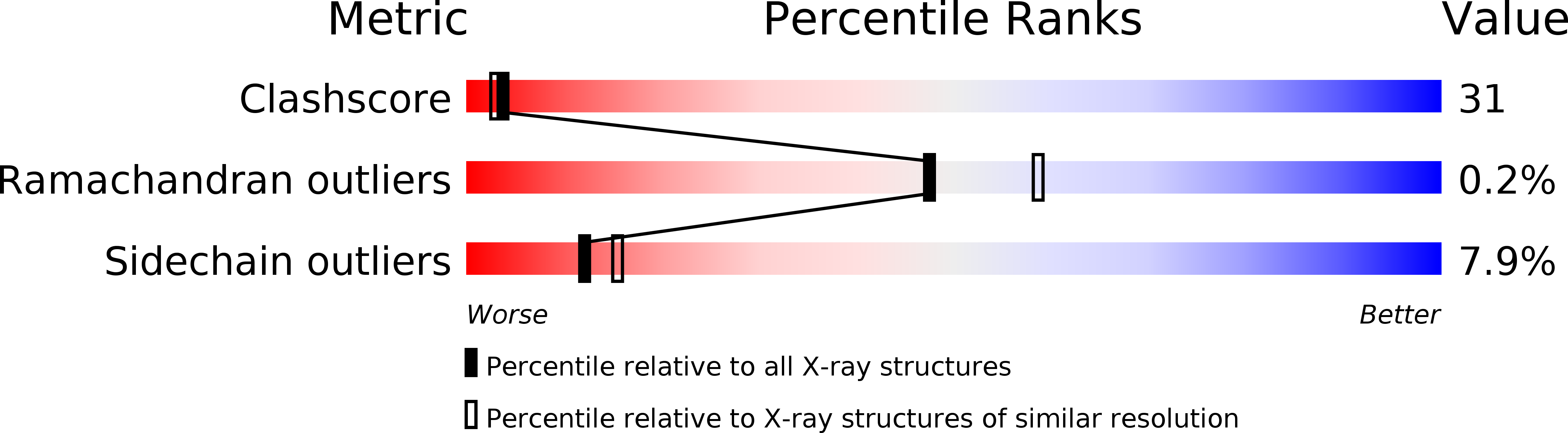
Experimental Data Snapshot
wwPDB Validation 3D Report Full Report
Entity ID: 1 | |||||
|---|---|---|---|---|---|
| Molecule | Chains | Sequence Length | Organism | Details | Image |
| Chitin Oligosaccharide Binding Protein | 529 | Vibrio cholerae | Mutation(s): 0 |  | |
UniProt | |||||
Find proteins for Q9KUA3 (Vibrio cholerae serotype O1 (strain ATCC 39315 / El Tor Inaba N16961)) Explore Q9KUA3 Go to UniProtKB: Q9KUA3 | |||||
Entity Groups | |||||
| Sequence Clusters | 30% Identity50% Identity70% Identity90% Identity95% Identity100% Identity | ||||
| UniProt Group | Q9KUA3 | ||||
Sequence AnnotationsExpand | |||||
| |||||
| Length ( Å ) | Angle ( ˚ ) |
|---|---|
| a = 92.813 | α = 90 |
| b = 92.813 | β = 90 |
| c = 137.029 | γ = 120 |
| Software Name | Purpose |
|---|---|
| DENZO | data reduction |
| SCALEPACK | data scaling |
| CNS | refinement |
| CNS | phasing |
RCSB PDB (citation) is hosted by
RCSB PDB is a member of the

