1ZDP

Crystal Structure Analysis of Thermolysin Complexed with the Inhibitor (S)-thiorphan


Experimental Data Snapshot

  • Method: X-RAY DIFFRACTION
  • Resolution: 1.70 Å
  • R-Value Work: 0.183 
  • R-Value Observed: 0.183 

wwPDB Validation   3D Report Full Report


This is version 1.4 of the entry. See complete history


Literature

Thiorphan and retro-thiorphan display equivalent interactions when bound to crystalline thermolysin

Roderick, S.L.Fournie-Zaluski, M.C.Roques, B.P.Matthews, B.W.

(1989) Biochemistry 28: 1493-1497

  • DOI: https://doi.org/10.1021/bi00430a011
  • Primary Citation of Related Structures:  
    1Z9G, 1ZDP

  • PubMed Abstract: 

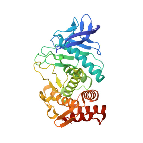
    The three-dimensional structures of (S)-thiorphan and (R)-retro-thiorphan bound to thermolysin have been determined crystallographically and refined to residuals of 0.183 and 0.187 at 1.7-A resolution. Thiorphan [N-[(S)-2-(mercaptomethyl)-1-oxo-3-phenylpropyl]glycine] [HSCH2CH(CH2C6H5)CONHC-H2COOH] and retro-thiorphan [[[(R)-1-(mercaptomethyl)-2-phenylethyl] amino]-3-oxopropanoic acid] [HSCH2CH(CH2C6H5)NHCOCH2COOH] are isomeric thiol-containing inhibitors of endopeptidase EC 24-11 (also called "enkephalinase"). The mode of binding of thiorphan to thermolysin is similar to that of (2-benzyl-3-mercaptopropanoyl)-L-alanylglycinamide [Monzingo, A.F., & Matthews, B.W. (1982) Biochemistry 21, 3390-3394] with the inhibitor sulfur atom coordinated to the active site zinc and the peptide portion forming substrate-like interactions with the enzyme. The isomeric inhibitor retro-thiorphan, which differs from thiorphan by the inversion of an amide bond, utilizes very similar interactions with enzyme. Despite the inversion of the -CO-NH- linkage the carbonyl oxygen and amide nitrogen display very similar hydrogen bonding, as anticipated by B.P. Roques et al. [(1983) Proc. Natl. Acad. Sci. U.S.A. 80, 3178-3182]. These results explain why thermolysin and possibly other zinc endopeptidases such as endopeptidase EC 24-11 fail to discriminate between these retro-inverso inhibitors.


  • Organizational Affiliation

    Institute of Molecular Biology, University of Oregon, Eugene 97403.


Macromolecules
Find similar proteins by:  (by identity cutoff)  |  3D Structure
Entity ID: 1
MoleculeChains Sequence LengthOrganismDetailsImage
ThermolysinA [auth E]316Bacillus thermoproteolyticusMutation(s): 0 
EC: 3.4.24.27
UniProt
Find proteins for P00800 (Bacillus thermoproteolyticus)
Explore P00800 
Go to UniProtKB:  P00800
Entity Groups  
Sequence Clusters30% Identity50% Identity70% Identity90% Identity95% Identity100% Identity
UniProt GroupP00800
Sequence Annotations
Expand
  • Reference Sequence
Small Molecules
Binding Affinity Annotations 
IDSourceBinding Affinity
TIO PDBBind:  1ZDP Ki: 1800 (nM) from 1 assay(s)
BindingDB:  1ZDP Ki: 1800 (nM) from 1 assay(s)
IC50: min: 1800, max: 9500 (nM) from 2 assay(s)
Binding MOAD:  1ZDP Ki: 1800 (nM) from 1 assay(s)
Experimental Data & Validation

Experimental Data

  • Method: X-RAY DIFFRACTION
  • Resolution: 1.70 Å
  • R-Value Work: 0.183 
  • R-Value Observed: 0.183 
  • Space Group: P 61 2 2
Unit Cell:
Length ( Å )Angle ( ˚ )
a = 94α = 90
b = 94β = 90
c = 132.1γ = 120
Software Package:
Software NamePurpose
OSCTSTdata collection
ROTAVATAdata reduction
TNTrefinement
OSCTSTdata reduction
CCP4data scaling
ROTAVATAdata scaling
TNTphasing

Structure Validation

View Full Validation Report


Entry History 

Deposition Data

Revision History  (Full details and data files)

  • Version 1.0: 2005-04-26
    Type: Initial release
  • Version 1.1: 2008-04-30
    Changes: Version format compliance
  • Version 1.2: 2011-07-13
    Changes: Version format compliance
  • Version 1.3: 2017-10-11
    Changes: Refinement description
  • Version 1.4: 2023-08-23
    Changes: Data collection, Database references, Derived calculations, Refinement description