Regulation by oligomerization in a mycobacterial folate biosynthetic enzyme.
Goulding, C.W., Apostol, M.I., Sawaya, M.R., Phillips, M., Parseghian, A., Eisenberg, D.(2005) J Mol Biol 349: 61-72
- PubMed: 15876368
- DOI: https://doi.org/10.1016/j.jmb.2005.03.023
- Primary Citation of Related Structures:
1NBU, 1Z9W - PubMed Abstract:
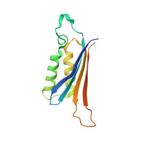
Folate derivatives are essential cofactors in the biosynthesis of purines, pyrimidines and amino acids across all forms of life. Mammals uptake folate from their diets, whereas most bacteria must synthesize folate de novo. Therefore, the enzymes in the folate biosynthetic pathway are attractive drug targets against bacterial pathogens such as Mycobacterium tuberculosis, the cause of the world's most deadly infectious disease, tuberculosis (TB). M.tuberculosis 7,8-dihydroneopterin aldolase (Mtb FolB, DHNA) is the second enzyme in the folate biosynthetic pathway, which catalyzes the conversion of 7,8-dihydroneopterin to 6-hydroxymethyl-7,8-dihydropterin and glycoaldehyde. The 1.6A X-ray crystal structure of Mtb FolB complexed with its product, 6-hydroxymethyl-7,8-dihydropterin, reveals an octameric assembly similar to that seen in crystal structures of other FolB homologs. However, the 2.5A crystal structure of unliganded Mtb FolB reveals a novel tetrameric oligomerization state, with only partially formed active sites. A substrate induced conformational change appears to be necessary to convert the inactive tetramer to the active octamer. Ultracentrifugation confirmed that in solution unliganded Mtb FolB is mainly tetrameric and upon addition of substrate FolB is predominantly octameric. Kinetic analysis of substrate binding gives a Hill coefficient of 2.0, indicating positive cooperativity. We hypothesize that Mtb FolB displays cooperativity in substrate binding to regulate the cellular concentration of 7,8-dihydroneopterin, so that it may function not only as a precursor to folate but also as an antioxidant for the survival of M.tuberculosis against host defenses.
Organizational Affiliation:
Molecular Biology Institute UCLA-DOE Institute of Genomics and Proteomics, P.O. Box 951570, Los Angeles, CA 90095-1570, USA.