Structure of HinP1I endonuclease reveals a striking similarity to the monomeric restriction enzyme MspI
Yang, Z., Horton, J.R., Maunus, R., Wilson, G.G., Roberts, R.J., Cheng, X.(2005) Nucleic Acids Res 33: 1892-1901
- PubMed: 15805123
- DOI: https://doi.org/10.1093/nar/gki337
- Primary Citation of Related Structures:
1YNM - PubMed Abstract:
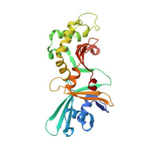
HinP1I, a type II restriction endonuclease, recognizes and cleaves a palindromic tetranucleotide sequence (G/CGC) in double-stranded DNA, producing 2 nt 5' overhanging ends. Here, we report the structure of HinP1I crystallized as one protein monomer in the crystallographic asymmetric unit. HinP1I displays an elongated shape, with a conserved catalytic core domain containing an active-site motif of SDX18QXK and a putative DNA-binding domain. Without significant sequence homology, HinP1I displays striking structural similarity to MspI, an endonuclease that cleaves a similar palindromic DNA sequence (C/CGG) and binds to that sequence crystallographically as a monomer. Almost all the structural elements of MspI can be matched in HinP1I, including both the DNA recognition and catalytic elements. Examining the protein-protein interactions in the crystal lattice, HinP1I could be dimerized through two helices located on the opposite side of the protein to the active site, generating a molecule with two active sites and two DNA-binding surfaces opposite one another on the outer surfaces of the dimer. A possible functional link between this unusual dimerization mode and the tetrameric restriction enzymes is discussed.
Organizational Affiliation:
Department of Biochemistry, Emory University School of Medicine 1510 Clifton Road, Atlanta, GA 30322, USA.