Triosephosphate isomerase from Plasmodium falciparum: the crystal structure provides insights into antimalarial drug design.
Velanker, S.S., Ray, S.S., Gokhale, R.S., Suma, S., Balaram, H., Balaram, P., Murthy, M.R.(1997) Structure 5: 751-761
- PubMed: 9261072
- DOI: https://doi.org/10.1016/s0969-2126(97)00230-x
- Primary Citation of Related Structures:
1YDV - PubMed Abstract:
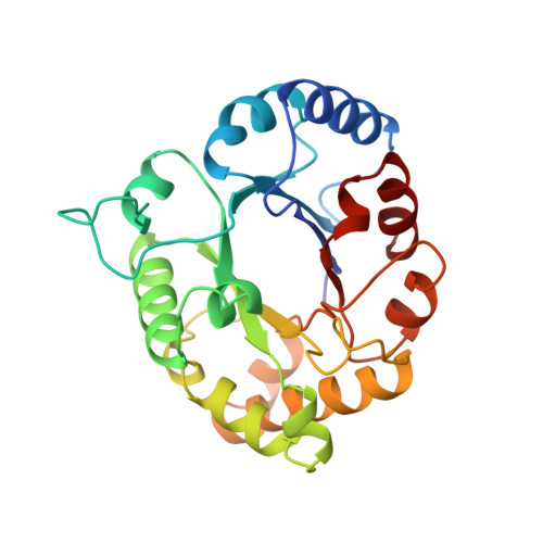
Malaria caused by the parasite Plasmodium falciparum is a major public health concern. The parasite lacks a functional tricarboxylic acid cycle, making glycolysis its sole energy source. Although parasite enzymes have been considered as potential antimalarial drug targets, little is known about their structural biology. Here we report the crystal structure of triosephosphate isomerase (TIM) from P. falciparum at 2.2 A resolution. The crystal structure of P. falciparum TIM (PfTIM), expressed in Escherichia coli, was determined by the molecular replacement method using the structure of trypanosomal TIM as the starting model. Comparison of the PfTIM structure with other TIM structures, particularly human TIM, revealed several differences. In most TIMs the residue at position 183 is a glutamate but in PfTIM it is a leucine. This leucine residue is completely exposed and together with the surrounding positively charged patch, may be responsible for binding TIM to the erythrocyte membrane. Another interesting feature is the occurrence of a cysteine residue at the dimer interface of PfTIM (Cys13), in contrast to human TIM where this residue is a methionine. Finally, residue 96 of human TIM (Ser96), which occurs near the active site, has been replaced by phenylalanine in PfTIM. Although the human and Plasmodium enzymes share 42% amino acid sequence identity, several key differences suggest that PfTIM may turn out to be a potential drug target. We have identified a region which may be responsible for binding PfTIM to cytoskeletal elements or the band 3 protein of erythrocytes; attachment to the erythrocyte membrane may subsequently lead to the extracellular exposure of parts of the protein. This feature may be important in view of a recent report that patients suffering from P. falciparum malaria mount an antibody response to TIM leading to prolonged hemolysis. A second approach to drug design may be provided by the mutation of the largely conserved residue (Ser96) to phenylalanine in PfTIM. This difference may be of importance in designing specific active-site inhibitors against the enzyme. Finally, specific inhibition of PfTIM subunit assembly might be possible by targeting Cys13 at the dimer interface. The crystal structure of PfTIM provides a framework for new therapeutic leads.
Organizational Affiliation:
Molecular Biophysics Unit, Indian Institute of Science, Bangalore, India.