1Y75

A new form of catalytically inactive phospholipase A2 with an unusual disulphide bridge Cys 32- Cys 49 reveals recognition for N-acetylglucosmine


Experimental Data Snapshot

  • Method: X-RAY DIFFRACTION
  • Resolution: 2.30 Å
  • R-Value Free: 0.256 
  • R-Value Work: 0.216 
  • R-Value Observed: 0.216 

wwPDB Validation   3D Report Full Report


This is version 1.3 of the entry. See complete history


Literature

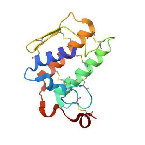
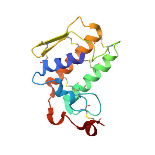
Crystal structure of a heterodimer of phospholipase A2 from Naja naja sagittifera at 2.3 A resolution reveals the presence of a new PLA2-like protein with a novel cys 32-Cys 49 disulphide bridge with a bound sugar at the substrate-binding site

Jabeen, T.Singh, N.Singh, R.K.Jasti, J.Sharma, S.Kaur, P.Srinivasan, A.Singh, T.P.

(2006) Proteins 62: 329-337

  • DOI: https://doi.org/10.1002/prot.20708
  • Primary Citation of Related Structures:  
    1Y75

  • PubMed Abstract: 

    The crystal structure of the phospholipase A2 (PLA2) heterodimer from Naja naja sagittifera reveals the presence of a new PLA2-like protein with eight disulphide bridges. The heterodimer is formed between a commonly observed group I PLA2 having seven characteristic disulfide bonds and a novel PLA2-like protein (Cys-PLA2) containing two extra cysteines at two highly conserved sites (positions 32 and 49) of structural and functional importance. The crystals of the heterodimer belong to tetragonal space group P41212 with cell dimensions, a = b = 77.7 A and c = 68.4 A corresponding to a solvent content of 33%, which is one of the lowest values observed so far in the PLA2 crystals. The structure has been solved with molecular replacement method and refined to a final R value of 21.6% [Rfree = 25.6%]. The electron density revealed the presence of cysteines 32 and 49 that are covalently linked to give rise to an eighth disulphide bridge in the PLA2-like monomer. A non-protein high-quality electron density was also observed at the substrate-binding site in the PLA2-like protein that has been interpreted as N-acetylglucosamine. The overall tertiary folds of the two monomers are similar having all features of PLA2-type folding. A zinc ion is detected at the interface of the heterodimer with fivefold coordination while another zinc ion was found on the surface of Cys-PLA2 with sixfold coordination. The conformations of the calcium-binding loops of both monomers are significantly different from each other as well as from those in other group I PLA2s. The N-acetylglucosamine molecule is favorably placed in the substrate-binding site of Cys-PLA2 and forms five hydrogen bonds and several van der Waals interactions with protein atoms, thus indicating a strong affinity. It also provides clue of the possible mechanism of sugar recognition by PLA2 and PLA2-like proteins. The formation of heterodimer seems to have been induced by zinc ion.


  • Organizational Affiliation

    Department of Biophysics, All India Institute of Medical Sciences, New Delhi, India.


Macromolecules
Find similar proteins by:  (by identity cutoff)  |  3D Structure
Entity ID: 1
MoleculeChains Sequence LengthOrganismDetailsImage
phospholipase A2 isoform 5118Naja sagittiferaMutation(s): 0 
EC: 3.1.1.4
Membrane Entity: Yes 
UniProt
Find proteins for Q5G291 (Naja sagittifera)
Explore Q5G291 
Go to UniProtKB:  Q5G291
Entity Groups  
Sequence Clusters30% Identity50% Identity70% Identity90% Identity95% Identity100% Identity
UniProt GroupQ5G291
Sequence Annotations
Expand
  • Reference Sequence
Find similar proteins by:  (by identity cutoff)  |  3D Structure
Entity ID: 2
MoleculeChains Sequence LengthOrganismDetailsImage
phospholipase A2 isoform 6118Naja sagittiferaMutation(s): 0 
EC: 3.1.1.4
Membrane Entity: Yes 
UniProt
Find proteins for Q5G290 (Naja sagittifera)
Explore Q5G290 
Go to UniProtKB:  Q5G290
Entity Groups  
Sequence Clusters30% Identity50% Identity70% Identity90% Identity95% Identity100% Identity
UniProt GroupQ5G290
Sequence Annotations
Expand
  • Reference Sequence
Small Molecules
Ligands 2 Unique
IDChains Name / Formula / InChI Key2D Diagram3D Interactions
NAG
Query on NAG

Download Ideal Coordinates CCD File 
C [auth A]2-acetamido-2-deoxy-beta-D-glucopyranose
C8 H15 N O6
OVRNDRQMDRJTHS-FMDGEEDCSA-N
ZN
Query on ZN

Download Ideal Coordinates CCD File 
D [auth A],
E [auth A]
ZINC ION
Zn
PTFCDOFLOPIGGS-UHFFFAOYSA-N
Experimental Data & Validation

Experimental Data

  • Method: X-RAY DIFFRACTION
  • Resolution: 2.30 Å
  • R-Value Free: 0.256 
  • R-Value Work: 0.216 
  • R-Value Observed: 0.216 
  • Space Group: P 41 21 2
Unit Cell:
Length ( Å )Angle ( ˚ )
a = 77.656α = 90
b = 77.656β = 90
c = 68.424γ = 90
Software Package:
Software NamePurpose
CNSrefinement
DENZOdata reduction
SCALEPACKdata scaling
AMoREphasing

Structure Validation

View Full Validation Report


Entry History 

Deposition Data

Revision History  (Full details and data files)

  • Version 1.0: 2005-05-03
    Type: Initial release
  • Version 1.1: 2008-04-30
    Changes: Version format compliance
  • Version 1.2: 2011-07-13
    Changes: Non-polymer description, Version format compliance
  • Version 1.3: 2020-07-29
    Type: Remediation
    Reason: Carbohydrate remediation
    Changes: Data collection, Derived calculations, Structure summary