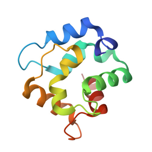
Crystal Structure of the D94S/G98E Variant of Rat alpha-Parvalbumin. An Explanation for the Reduced Divalent Ion Affinity.
Tanner, J.J., Agah, S., Lee, Y.H., Henzl, M.T.(2005) Biochemistry 44: 10966-10976
- PubMed: 16101280
- DOI: https://doi.org/10.1021/bi050770t
- Primary Citation of Related Structures:
1XVJ - PubMed Abstract:
Simultaneous replacement of Asp-94 with serine and Gly-98 with glutamate in rat alpha-parvalbumin creates a CD-site ligand array in the context of the EF-site binding loop. Previous work has shown that, relative to the wild-type CD site, this engineered site has markedly reduced Ca(2+) affinity. Seeking an explanation for this phenomenon, we have obtained the crystal structure of the alpha D94S/G98E variant. The Ca(2+) coordination within the engineered EF site of the 94/98E variant is nearly identical to that within the CD site, suggesting that the attenuated affinity of the EF site in 94/98E is not a consequence of suboptimal coordination geometry. We have also examined the divalent ion binding properties of the alpha 94/98E variant in both Na(+)- and K(+)-containing buffers. Although the Ca(2+) and Mg(2+) affinities are higher in K(+) solution, the increases are comparable to those observed for wild-type alpha. Consistent with that finding, the apparent Na(+) stoichiometry, estimated from stability studies conducted as a function of Na(+) concentration, is 1.0 +/- 0.1, identical to that of wild-type alpha. Thus, the reduced affinity for divalent ions is evidently not the result of heightened monovalent ion competition. The thermodynamic analysis indicates that the less favorable Gibbs free energy of binding reflects a substantial enthalpic penalty. Significantly, the crystal structure reveals a steric clash between Phe-57 and the C(gamma) atom of Glu-98. The consequent displacement of Phe-57 also produces a close contact with Ser-55. Thus, steric interference may be the source of the enthalpic penalty.
Organizational Affiliation:
Department of Chemistry, University of Missouri, Columbia, Missouri 65211, USA.