1XDK

Crystal Structure of the RARbeta/RXRalpha Ligand Binding Domain Heterodimer in Complex with 9-cis Retinoic Acid and a Fragment of the TRAP220 Coactivator


Experimental Data Snapshot

  • Method: X-RAY DIFFRACTION
  • Resolution: 2.90 Å
  • R-Value Free: 0.296 
  • R-Value Work: 0.253 
  • R-Value Observed: 0.253 

wwPDB Validation   3D Report Full Report


Ligand Structure Quality Assessment 


This is version 1.5 of the entry. See complete history


Literature

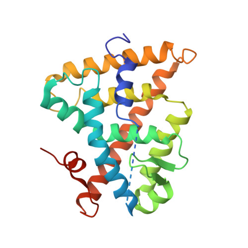
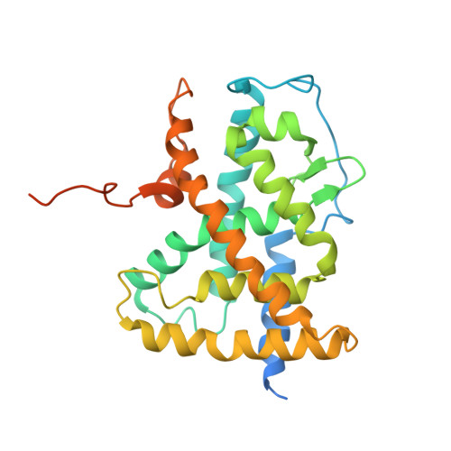
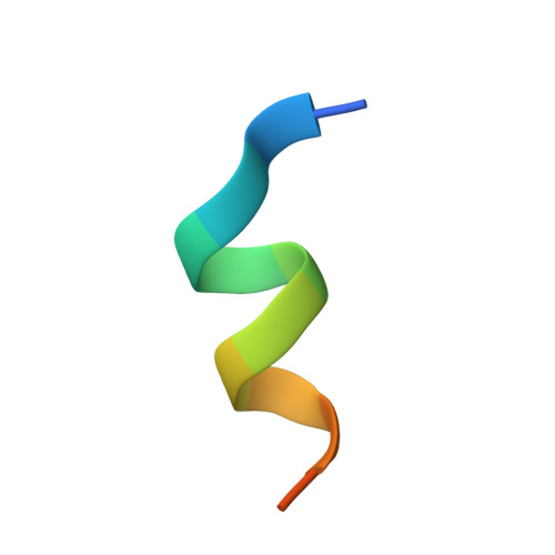
CHARACTERIZATION OF THE INTERACTION BETWEEN RAR/RXR HETERODIMERS AND TRANSCRIPTIONAL COACTIVATORS THROUGH STRUCTURAL AND FLUORESCENCE ANISOTROPY STUDIES

Pogenberg, V.Guichou, J.F.Vivat-Hannah, V.Kammerer, S.Perez, E.Germain, P.De Lera, A.R.Gronemeyer, H.Royer, C.A.Bourguet, W.

(2005) J Biol Chem 280: 1625-1633

  • DOI: https://doi.org/10.1074/jbc.M409302200
  • Primary Citation of Related Structures:  
    1XDK

  • PubMed Abstract: 

    Retinoid receptors (RARs and RXRs) are ligand-activated transcription factors that regulate the transcription of target genes by recruiting coregulator complexes at cognate promoters. To understand the effects of heterodimerization and ligand binding on coactivator recruitment, we solved the crystal structure of the complex between the RARbeta/RXRalpha ligand-binding domain heterodimer, its 9-cis retinoic acid ligand, and an LXXLL-containing peptide (termed NR box 2) derived from the nuclear receptor interaction domain (NID) of the TRAP220 coactivator. In parallel, we measured the binding affinities of the isolated NR box 2 peptide or the full-length NID of the coactivator SRC-1 for retinoid receptors in the presence of various types of ligands. Our correlative analysis of three-dimensional structures and fluorescence data reveals that heterodimerization does not significantly alter the structure of individual subunits or their intrinsic capacity to interact with NR box 2. Similarly, we show that the ability of a protomer to recruit NR box 2 does not vary as a function of the ligand binding status of the partner receptor. In contrast, the strength of the overall association between the heterodimer and the full-length SRC-1 NID is dictated by the combinatorial action of RAR and RXR ligands, the simultaneous presence of the two receptor agonists being required for highest binding affinity. We identified an LXXLL peptide-driven mechanism by which the concerted reorientation of three phenylalanine side chains generates an "aromatic clamp" that locks the RXR activation helix H12 in the transcriptionally active conformation. Finally, we show how variations of helix H11-ligand interactions can alter the communication pathway linking helices H11, H12, and the connecting loop L11-12 to the coactivator-binding site. Together, our results reveal molecular and structural features that impact on the ligand-dependent interaction of the RAR/RXR heterodimer with nuclear receptor coactivators.


  • Organizational Affiliation

    Centre de Biochimie Structurale, CNRS U5048-INSERM U554-UM1, Faculté de Pharmacie, 15 avenue Charles Flahault, 34093 Montpellier, France.


Macromolecules
Find similar proteins by:  (by identity cutoff)  |  3D Structure
Entity ID: 1
MoleculeChains Sequence LengthOrganismDetailsImage
Retinoic acid receptor RXR-alpha
A, E
238Mus musculusMutation(s): 0 
Gene Names: RxraNr2b1
UniProt
Find proteins for P28700 (Mus musculus)
Explore P28700 
Go to UniProtKB:  P28700
Entity Groups  
Sequence Clusters30% Identity50% Identity70% Identity90% Identity95% Identity100% Identity
UniProt GroupP28700
Sequence Annotations
Expand
  • Reference Sequence
Find similar proteins by:  (by identity cutoff)  |  3D Structure
Entity ID: 2
MoleculeChains Sequence LengthOrganismDetailsImage
Retinoic acid receptor, beta
B, F
303Mus musculusMutation(s): 0 
Gene Names: RarbNr1b2
UniProt
Find proteins for P22605 (Mus musculus)
Explore P22605 
Go to UniProtKB:  P22605
Entity Groups  
Sequence Clusters30% Identity50% Identity70% Identity90% Identity95% Identity100% Identity
UniProt GroupP22605
Sequence Annotations
Expand
  • Reference Sequence

Find similar proteins by:  Sequence   |   3D Structure  

Entity ID: 3
MoleculeChains Sequence LengthOrganismDetailsImage
Thyroid Receptor Associated Protein 220
C, D, G, H
14N/AMutation(s): 0 
UniProt & NIH Common Fund Data Resources
Find proteins for Q925J9 (Mus musculus)
Explore Q925J9 
Go to UniProtKB:  Q925J9
IMPC:  MGI:1100846
Entity Groups  
Sequence Clusters30% Identity50% Identity70% Identity90% Identity95% Identity100% Identity
UniProt GroupQ925J9
Sequence Annotations
Expand
  • Reference Sequence
Small Molecules
Binding Affinity Annotations 
IDSourceBinding Affinity
9CR BindingDB:  1XDK Ki: min: 0.5, max: 583 (nM) from 16 assay(s)
Kd: min: 0.2, max: 1810 (nM) from 29 assay(s)
IC50: min: 4, max: 82 (nM) from 8 assay(s)
EC50: min: 1.5, max: 316 (nM) from 34 assay(s)
Experimental Data & Validation

Experimental Data

  • Method: X-RAY DIFFRACTION
  • Resolution: 2.90 Å
  • R-Value Free: 0.296 
  • R-Value Work: 0.253 
  • R-Value Observed: 0.253 
  • Space Group: P 31 2 1
Unit Cell:
Length ( Å )Angle ( ˚ )
a = 115.7α = 90
b = 115.7β = 90
c = 247.2γ = 120
Software Package:
Software NamePurpose
MAR345data collection
SCALAdata scaling
MOLREPphasing
CNSrefinement
MOSFLMdata reduction
CCP4data scaling

Structure Validation

View Full Validation Report


Ligand Structure Quality Assessment 


Entry History 

Deposition Data

Revision History  (Full details and data files)

  • Version 1.0: 2004-11-09
    Type: Initial release
  • Version 1.1: 2008-04-30
    Changes: Version format compliance
  • Version 1.2: 2011-07-13
    Changes: Version format compliance
  • Version 1.3: 2016-11-16
    Changes: Non-polymer description
  • Version 1.4: 2017-10-11
    Changes: Refinement description
  • Version 1.5: 2023-08-23
    Changes: Data collection, Database references, Derived calculations, Refinement description