Structure of a mutant T=1 capsid of Sesbania mosaic virus: role of water molecules in capsid architecture and integrity.
Sangita, V., Satheshkumar, P.S., Savithri, H.S., Murthy, M.R.(2005) Acta Crystallogr D Biol Crystallogr 61: 1406-1412
- PubMed: 16204894
- DOI: https://doi.org/10.1107/S0907444905024030
- Primary Citation of Related Structures:
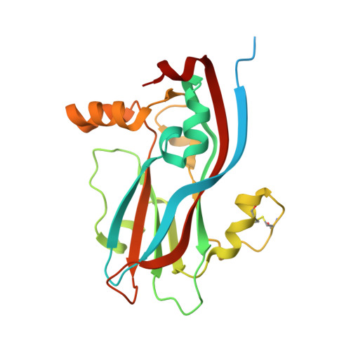
1X36 - PubMed Abstract:
Deletion of the N-terminal 31 amino acids from the coat protein (CP) of Sesbania mosaic virus (SeMV) results in the formation of T = 1 capsids. The X-ray crystal structure of CP-NDelta31 mutant capsids reveals that the CP adopts a conformation similar to those of other T = 1 mutants. The 40 N-terminal residues are disordered in CP-NDelta31. The intersubunit hydrogen bonds closely resemble those of the native capsid. The role of water molecules in the SeMV structure has been analyzed for the first time using the present structure. As many as 139 of the 173 waters per subunit make direct contacts with the protein atoms. The water molecules form a robust scaffold around the capsid, stabilize the loops and provide integrity to the subunit. These waters constitute a network connecting diametrically opposite ends of the subunit. Such waters might act as nodes for conveying signals for assembly or disassembly across a large conformational space. Many water-mediated interactions are observed at various interfaces. The twofold interface, which has the smallest number of protein-protein contacts, is primarily held by water-mediated interactions. The present structure illuminates the role of water molecules in the structure and stability of the capsid and points out their possible significance in assembly.
Organizational Affiliation:
Molecular Biophysics Unit, Indian Institute of Science, Bangalore 560012, India.