1WZU

Crystal structure of quinolinate synthase (nadA)


Experimental Data Snapshot

  • Method: X-RAY DIFFRACTION
  • Resolution: 2.00 Å
  • R-Value Free: 0.268 
  • R-Value Work: 0.217 
  • R-Value Observed: 0.222 

wwPDB Validation   3D Report Full Report


This is version 1.3 of the entry. See complete history


Literature

Crystal Structure of the NAD Biosynthetic Enzyme Quinolinate Synthase

Sakuraba, H.Tsuge, H.Yoneda, K.Katunuma, N.Ohshima, T.

(2005) J Biol Chem 280: 26645-26648

  • DOI: https://doi.org/10.1074/jbc.C500192200
  • Primary Citation of Related Structures:  
    1WZU

  • PubMed Abstract: 

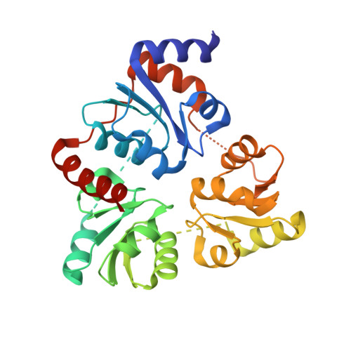
    A gene encoding a quinolinate synthase has been identified in the hyperthermophilic archaeon Pyrococcus horikoshii via genome sequencing. The gene was overexpressed in Escherichia coli, and the crystal structure of the produced enzyme was determined to 2.0 A resolution in the presence of malate, a substrate analogue. The overall structure exhibits a unique triangular architecture composed of a 3-fold repeat of three-layer (alphabetaalpha) sandwich folding. Although some aspects of the fold homologous to the each domain have been observed previously, the overall structure of quinolinate synthase shows no similarity to any known protein structure. The three analogous domains are related to a pseudo-3-fold symmetry. The active site is located at the interface of the three domains and is centered on the pseudo-3-fold axis. The malate molecule is tightly held near the bottom of the active site cavity. The model of the catalytic state during the first condensation step of the quinolinate synthase reaction indicates that the elimination of inorganic phosphate from dihydroxyacetone phosphate may precede the condensation reaction.


  • Organizational Affiliation

    Department of Biological Science and Technology, Faculty of Engineering, The University of Tokushima, 2-1 Minamijosanjima-cho, Tokushima 770-8506.


Macromolecules
Find similar proteins by:  (by identity cutoff)  |  3D Structure
Entity ID: 1
MoleculeChains Sequence LengthOrganismDetailsImage
Quinolinate synthetase A300Pyrococcus horikoshiiMutation(s): 0 
UniProt
Find proteins for O57767 (Pyrococcus horikoshii (strain ATCC 700860 / DSM 12428 / JCM 9974 / NBRC 100139 / OT-3))
Explore O57767 
Go to UniProtKB:  O57767
Entity Groups  
Sequence Clusters30% Identity50% Identity70% Identity90% Identity95% Identity100% Identity
UniProt GroupO57767
Sequence Annotations
Expand
  • Reference Sequence
Small Molecules
Ligands 1 Unique
IDChains Name / Formula / InChI Key2D Diagram3D Interactions
MLT
Query on MLT

Download Ideal Coordinates CCD File 
B [auth A]D-MALATE
C4 H6 O5
BJEPYKJPYRNKOW-UWTATZPHSA-N
Experimental Data & Validation

Experimental Data

  • Method: X-RAY DIFFRACTION
  • Resolution: 2.00 Å
  • R-Value Free: 0.268 
  • R-Value Work: 0.217 
  • R-Value Observed: 0.222 
  • Space Group: P 63
Unit Cell:
Length ( Å )Angle ( ˚ )
a = 94.61α = 90
b = 94.61β = 90
c = 62.57γ = 120
Software Package:
Software NamePurpose
HKL-2000data collection
SCALEPACKdata scaling
SOLVEphasing
CNSrefinement
HKL-2000data reduction

Structure Validation

View Full Validation Report


Entry History 

Deposition Data

  • Released Date: 2005-06-07 
  • Deposition Author(s): Sakuraba, H.

Revision History  (Full details and data files)

  • Version 1.0: 2005-06-07
    Type: Initial release
  • Version 1.1: 2008-04-30
    Changes: Version format compliance
  • Version 1.2: 2011-07-13
    Changes: Version format compliance
  • Version 1.3: 2024-03-13
    Changes: Data collection, Database references, Derived calculations