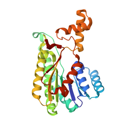
d-3-Hydroxybutyrate Dehydrogenase from Pseudomonas fragi: Molecular Cloning of the Enzyme Gene and Crystal Structure of the Enzyme
Ito, K., Nakajima, Y., Ichihara, E., Ogawa, K., Katayama, N., Nakashima, K., Yoshimoto, T.(2006) J Mol Biol 355: 722-733
- PubMed: 16325199
- DOI: https://doi.org/10.1016/j.jmb.2005.10.072
- Primary Citation of Related Structures:
1WMB, 1X1T - PubMed Abstract:
The gene coding for d-3-hydroxybutyrate dehydrogenase (HBDH) was cloned from Pseudomonas fragi. The nucleotide sequence contained a 780 bp open reading frame encoding a 260 amino acid residue protein. The recombinant enzyme was efficiently expressed in Escherichia coli cells harboring pHBDH11 and was purified to homogeneity as judged by SDS-PAGE. The enzyme showed a strict stereospecificity to the D-enantiomer (3R-configuration) of 3-hydroxybutyrate as a substrate. Crystals of the ligand-free HBDH and of the enzyme-NAD+ complex were obtained using the hanging-drop, vapor-diffusion method. The crystal structure of the HBDH was solved by the multiwavelength anomalous diffraction method using the SeMet-substituted enzyme and was refined to 2.0 A resolution. The overall structure of P.fragi HBDH, including the catalytic tetrad of Asn114, Ser142, Tyr155, and Lys159, shows obvious relationships with other members of the short-chain dehydrogenase/reductase (SDR) family. A cacodylate anion was observed in both the ligand-free enzyme and the enzyme-NAD+ complex, and was located near the catalytic tetrad. It was shown that the cacodylate inhibited the NAD+-dependent D-3-hydroxybutyrate dehydrogenation competitively, with a Ki value of 5.6 mM. From the interactions between cacodylate and the enzyme, it is predicted that substrate specificity is achieved through the recognition of the 3-methyl and carboxyl groups of the substrate.
Organizational Affiliation:
Department of Molecular Medicinal Sciences, Graduate School of Biomedical Sciences, Nagasaki University, 1-14 Bunkyo-machi, Nagasaki 852-8521, Japan. k.ito@net.nagasaki-u.ac.jp