


Crystal Structure of Alpha-Xylosidase from Escherichia coli
Ose, T., Kitamura, M., Okuyama, M., Mori, H., Kimura, A., Watanabe, N., Yao, M., Tanaka, I.To be published.
Experimental Data Snapshot
wwPDB Validation 3D Report Full Report

Entity ID: 1 | |||||
|---|---|---|---|---|---|
| Molecule | Chains | Sequence Length | Organism | Details | Image |
| Putative family 31 glucosidase yicI | 772 | Escherichia coli | Mutation(s): 19 Gene Names: YicI EC: 3.2.1 | 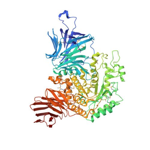 | |
UniProt | |||||
Find proteins for P31434 (Escherichia coli (strain K12)) Explore P31434 Go to UniProtKB: P31434 | |||||
Entity Groups | |||||
| Sequence Clusters | 30% Identity50% Identity70% Identity90% Identity95% Identity100% Identity | ||||
| UniProt Group | P31434 | ||||
Sequence AnnotationsExpand | |||||
| |||||
| Ligands 1 Unique | |||||
|---|---|---|---|---|---|
| ID | Chains | Name / Formula / InChI Key | 2D Diagram | 3D Interactions | |
| MES Query on MES | G [auth A] H [auth B] I [auth C] J [auth D] K [auth E] | 2-(N-MORPHOLINO)-ETHANESULFONIC ACID C6 H13 N O4 S SXGZJKUKBWWHRA-UHFFFAOYSA-N |  | ||
| Modified Residues 1 Unique | |||||
|---|---|---|---|---|---|
| ID | Chains | Type | Formula | 2D Diagram | Parent |
| MSE Query on MSE | A, B, C, D, E A, B, C, D, E, F | L-PEPTIDE LINKING | C5 H11 N O2 Se |  | MET |
| Length ( Å ) | Angle ( ˚ ) |
|---|---|
| a = 161.337 | α = 90 |
| b = 174.791 | β = 90 |
| c = 209.629 | γ = 90 |
| Software Name | Purpose |
|---|---|
| DENZO | data reduction |
| SCALEPACK | data scaling |
| SHARP | phasing |
| CNS | refinement |
| SnB | phasing |
| DM | phasing |
RCSB PDB (citation) is hosted by
RCSB PDB is a member of the

