1WDC

SCALLOP MYOSIN REGULATORY DOMAIN


Experimental Data Snapshot

  • Method: X-RAY DIFFRACTION
  • Resolution: 2.00 Å
  • R-Value Free: 0.281 
  • R-Value Work: 0.194 
  • R-Value Observed: 0.194 

wwPDB Validation   3D Report Full Report


This is version 1.3 of the entry. See complete history


Literature

Structure of the regulatory domain of scallop myosin at 2 A resolution: implications for regulation.

Houdusse, A.Cohen, C.

(1996) Structure 4: 21-32

  • DOI: https://doi.org/10.1016/s0969-2126(96)00006-8
  • Primary Citation of Related Structures:  
    1WDC

  • PubMed Abstract: 

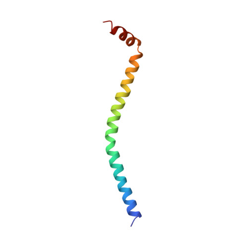
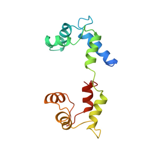
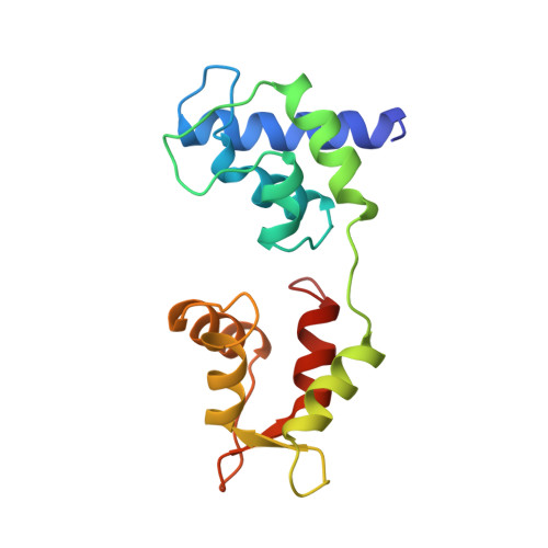
    In contrast to the myosins of vertebrate skeletal muscle, molluscan myosins are regulated molecules whose enzymatic activity is switched on by the direct binding of Ca2+. The head portion (S1) of the molecule consists of a motor domain and a regulatory domain (RD) containing a 'regulatory' and an 'essential' light chain (RLC and ELC, respectively). The structures of scallop myosin RD with bound Ca2+, as well as the S1 fragment of chicken skeletal muscle myosin, have been determined previously to 2.8 A resolution. We have determined the structure at 2.0 A resolution of scallop myosin RD with bound Ca2+. The unusual coordination at the specific Ca(2+)-binding site in the ELC has now been clarified, as has the structural basis for Mg2+ binding to the RLC. A comparison of the scallop RD structure with that in the chicken S1 structure shows differences in the bending of the two RDs in two different places. Based on these structural results, a model for regulation is proposed in which the Ca(2+)-bound RD is a rigid structure, and transient flexibility of the Ca(2+)-free RD allows the myosin heads to make stabilizing intramolecular linkage which shut off the motor.


  • Organizational Affiliation

    Rosenstiel Basic Medical Sciences Research Center, Brandeis University, Waltham, MA 02254-9110, USA.


Macromolecules
Find similar proteins by:  (by identity cutoff)  |  3D Structure
Entity ID: 1
MoleculeChains Sequence LengthOrganismDetailsImage
SCALLOP MYOSIN64Argopecten irradiansMutation(s): 0 
UniProt
Find proteins for P24733 (Argopecten irradians)
Explore P24733 
Go to UniProtKB:  P24733
Entity Groups  
Sequence Clusters30% Identity50% Identity70% Identity90% Identity95% Identity100% Identity
UniProt GroupP24733
Sequence Annotations
Expand
  • Reference Sequence
Find similar proteins by:  (by identity cutoff)  |  3D Structure
Entity ID: 2
MoleculeChains Sequence LengthOrganismDetailsImage
SCALLOP MYOSIN156Argopecten irradiansMutation(s): 0 
UniProt
Find proteins for P13543 (Argopecten irradians)
Explore P13543 
Go to UniProtKB:  P13543
Entity Groups  
Sequence Clusters30% Identity50% Identity70% Identity90% Identity95% Identity100% Identity
UniProt GroupP13543
Sequence Annotations
Expand
  • Reference Sequence
Find similar proteins by:  (by identity cutoff)  |  3D Structure
Entity ID: 3
MoleculeChains Sequence LengthOrganismDetailsImage
SCALLOP MYOSIN156Argopecten irradiansMutation(s): 0 
UniProt
Find proteins for P07291 (Argopecten irradians)
Explore P07291 
Go to UniProtKB:  P07291
Entity Groups  
Sequence Clusters30% Identity50% Identity70% Identity90% Identity95% Identity100% Identity
UniProt GroupP07291
Sequence Annotations
Expand
  • Reference Sequence
Small Molecules
Ligands 2 Unique
IDChains Name / Formula / InChI Key2D Diagram3D Interactions
CA
Query on CA

Download Ideal Coordinates CCD File 
E [auth C]CALCIUM ION
Ca
BHPQYMZQTOCNFJ-UHFFFAOYSA-N
MG
Query on MG

Download Ideal Coordinates CCD File 
D [auth B]MAGNESIUM ION
Mg
JLVVSXFLKOJNIY-UHFFFAOYSA-N
Experimental Data & Validation

Experimental Data

  • Method: X-RAY DIFFRACTION
  • Resolution: 2.00 Å
  • R-Value Free: 0.281 
  • R-Value Work: 0.194 
  • R-Value Observed: 0.194 
  • Space Group: P 1 21 1
Unit Cell:
Length ( Å )Angle ( ˚ )
a = 52.65α = 90
b = 87.1β = 114.5
c = 55.5γ = 90
Software Package:
Software NamePurpose
X-PLORmodel building
X-PLORrefinement
R-AXISdata reduction
CCP4data reduction
X-PLORphasing

Structure Validation

View Full Validation Report


Entry History 

Deposition Data

Revision History  (Full details and data files)

  • Version 1.0: 1996-07-11
    Type: Initial release
  • Version 1.1: 2008-03-24
    Changes: Version format compliance
  • Version 1.2: 2011-07-13
    Changes: Version format compliance
  • Version 1.3: 2024-02-14
    Changes: Data collection, Database references, Derived calculations, Other