1VST

Symmetric Sulfolobus solfataricus uracil phosphoribosyltransferase with bound PRPP and GTP


Experimental Data Snapshot

  • Method: X-RAY DIFFRACTION
  • Resolution: 2.80 Å
  • R-Value Free: 0.277 
  • R-Value Work: 0.226 

wwPDB Validation   3D Report Full Report


Ligand Structure Quality Assessment 


This is version 1.3 of the entry. See complete history


Literature

Structural and kinetic studies of the allosteric transition in Sulfolobus solfataricus uracil phosphoribosyltransferase: Permanent activation by engineering of the C-terminus

Christoffersen, S.Kadziola, A.Johansson, E.Rasmussen, M.Willemoes, M.Jensen, K.F.

(2009) J Mol Biol 393: 464-477

  • DOI: https://doi.org/10.1016/j.jmb.2009.08.019
  • Primary Citation of Related Structures:  
    1VST, 3G6W

  • PubMed Abstract: 

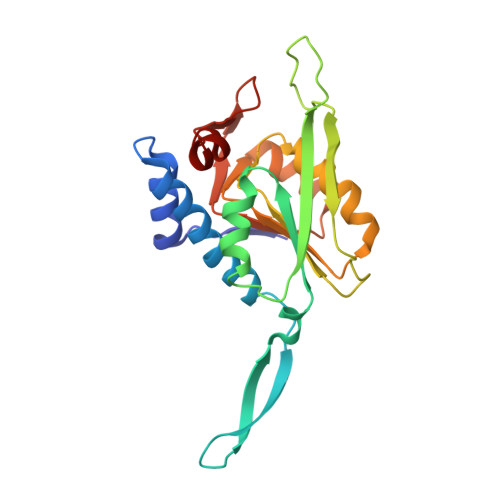
    Uracil phosphoribosyltransferase catalyzes the conversion of 5-phosphoribosyl-alpha-1-diphosphate (PRPP) and uracil to uridine monophosphate (UMP) and diphosphate (PP(i)). The tetrameric enzyme from Sulfolobus solfataricus has a unique type of allosteric regulation by cytidine triphosphate (CTP) and guanosine triphosphate (GTP). Here we report two structures of the activated state in complex with GTP. One structure (refined at 2.8-A resolution) contains PRPP in all active sites, while the other structure (refined at 2.9-A resolution) has PRPP in two sites and the hydrolysis products, ribose-5-phosphate and PP(i), in the other sites. Combined with three existing structures of uracil phosphoribosyltransferase in complex with UMP and the allosteric inhibitor cytidine triphosphate (CTP), these structures provide valuable insight into the mechanism of allosteric transition from inhibited to active enzyme. The regulatory triphosphates bind at a site in the center of the tetramer in a different manner and change the quaternary arrangement. Both effectors contact Pro94 at the beginning of a long beta-strand in the dimer interface, which extends into a flexible loop over the active site. In the GTP-bound state, two flexible loop residues, Tyr123 and Lys125, bind the PP(i) moiety of PRPP in the neighboring subunit and contribute to catalysis, while in the inhibited state, they contribute to the configuration of the active site for UMP rather than PRPP binding. The C-terminal Gly216 participates in a hydrogen-bond network in the dimer interface that stabilizes the inhibited, but not the activated, state. Tagging the C-terminus with additional amino acids generates an endogenously activated enzyme that binds GTP without effects on activity.


  • Organizational Affiliation

    Department of Biology, University of Copenhagen, Biocenter, Copenhagen N, Denmark.


Macromolecules
Find similar proteins by:  (by identity cutoff)  |  3D Structure
Entity ID: 1
MoleculeChains Sequence LengthOrganismDetailsImage
Uracil phosphoribosyltransferase216Saccharolobus solfataricusMutation(s): 0 
Gene Names: SSO0231upp
EC: 2.4.2.9
UniProt
Find proteins for Q980Q4 (Saccharolobus solfataricus (strain ATCC 35092 / DSM 1617 / JCM 11322 / P2))
Explore Q980Q4 
Go to UniProtKB:  Q980Q4
Entity Groups  
Sequence Clusters30% Identity50% Identity70% Identity90% Identity95% Identity100% Identity
UniProt GroupQ980Q4
Sequence Annotations
Expand
  • Reference Sequence
Small Molecules
Ligands 3 Unique
IDChains Name / Formula / InChI Key2D Diagram3D Interactions
GTP
Query on GTP

Download Ideal Coordinates CCD File 
B [auth A]GUANOSINE-5'-TRIPHOSPHATE
C10 H16 N5 O14 P3
XKMLYUALXHKNFT-UUOKFMHZSA-N
PRP
Query on PRP

Download Ideal Coordinates CCD File 
D [auth A]1-O-pyrophosphono-5-O-phosphono-alpha-D-ribofuranose
C5 H13 O14 P3
PQGCEDQWHSBAJP-TXICZTDVSA-N
MG
Query on MG

Download Ideal Coordinates CCD File 
C [auth A]MAGNESIUM ION
Mg
JLVVSXFLKOJNIY-UHFFFAOYSA-N
Experimental Data & Validation

Experimental Data

  • Method: X-RAY DIFFRACTION
  • Resolution: 2.80 Å
  • R-Value Free: 0.277 
  • R-Value Work: 0.226 
  • Space Group: P 64 2 2
Unit Cell:
Length ( Å )Angle ( ˚ )
a = 122.2α = 90
b = 122.2β = 90
c = 62.2γ = 120
Software Package:
Software NamePurpose
MAR345dtbdata collection
AMoREphasing
CNSrefinement
DENZOdata reduction
SCALEPACKdata scaling

Structure Validation

View Full Validation Report


Ligand Structure Quality Assessment 


Entry History 

Deposition Data

  • Released Date: 2009-09-01 
  • Deposition Author(s): Kadziola, A.

Revision History  (Full details and data files)

  • Version 1.0: 2009-09-01
    Type: Initial release
  • Version 1.1: 2011-07-13
    Changes: Version format compliance
  • Version 1.2: 2020-07-29
    Type: Remediation
    Reason: Carbohydrate remediation
    Changes: Data collection, Derived calculations, Structure summary
  • Version 1.3: 2023-10-25
    Changes: Data collection, Database references, Refinement description, Structure summary