Calf spleen purine-nucleoside phosphorylase: crystal structure of the binary complex with a potent multisubstrate analogue inhibitor.
Luic, M., Koellner, G., Yokomatsu, T., Shibuya, S., Bzowska, A.(2004) Acta Crystallogr D Biol Crystallogr 60: 1417-1424
- PubMed: 15272165
- DOI: https://doi.org/10.1107/S0907444904013861
- Primary Citation of Related Structures:
1V48 - PubMed Abstract:
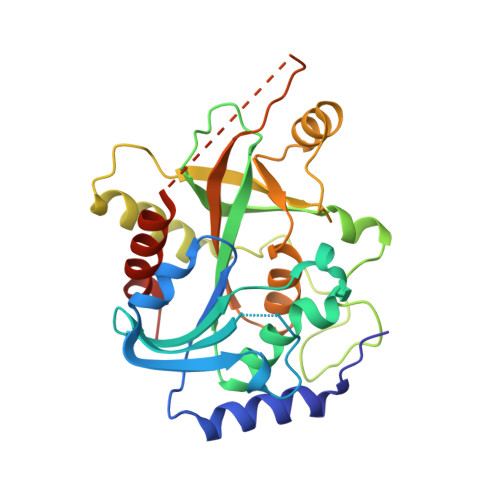
Purine-nucleoside phosphorylase (PNP) deficiency in humans leads to inhibition of the T-cell response. Potent membrane-permeable inhibitors of this enzyme are therefore considered to be potential immunosuppressive agents. The binary complex of the trimeric calf spleen phosphorylase, which is highly homologous to human PNP, with the potent ground-state analogue inhibitor 9-(5,5-difluoro-5-phosphonopentyl)guanine (DFPP-G) was crystallized in the cubic space group P2(1)3, with unit-cell parameter a = 93.183 A and one monomer per asymmetric unit. High-resolution X-ray diffraction data were collected using synchrotron radiation (EMBL Outstation, DESY, Hamburg, station X13). The crystal structure was refined to a resolution of 2.2 A and R and Rfree values of 19.1 and 24.2%, respectively. The crystal structure confirms that DFPP-G acts as a multisubstrate analogue inhibitor as it binds to both nucleoside- and phosphate-binding sites. The structure also provides the answers to some questions regarding the substrate specificity and molecular mechanism of trimeric PNPs. The wide access to the active-site pocket that was observed in the reported structure as a result of the flexibility or disorder of two loops (residues 60-65 and 251-266) strongly supports the random binding of substrates. The putative hydrogen bonds identified in the base-binding site indicate that N1-H and not O6 of the purine base defines the specificity of trimeric PNPs. This is confirmed by the fact that the contact of guanine O6 with Asn243 Odelta1 is not a direct contact but is mediated by a water molecule. Participation of Arg84 in the binding of the phosphonate group experimentally verifies the previous suggestion [Blackburn & Kent (1986), J. Chem. Soc. Perkin Trans. I, pp. 913-917; Halazy et al. (1991), J. Am. Chem. Soc. 113, 315-317] that fluorination of alkylphosphonates yields compounds with properties that suitably resemble those of phosphate esters and in turn leads to optimized interactions of such analogues with the phosphate-binding site residues. DFPP-G shows a Ki(app) in the nanomolar range towards calf and human PNPs. To date, no high-resolution X-ray structures of these enzymes with such potent ground-state analogue inhibitors have been available in the Protein Data Bank. The present structure may thus be used in the rational structure-based design of new PNP inhibitors with potential medical applications.
Organizational Affiliation:
Rudjer Bosković Institute, Bijenicka 54, 10002 Zagreb, Croatia.