


Crystal structure of a hypothetical protein yodA
Eswaramoorthy, S., Swaminathan, S.To be published.
Experimental Data Snapshot
wwPDB Validation 3D Report Full Report

Entity ID: 1 | |||||
|---|---|---|---|---|---|
| Molecule | Chains | Sequence Length | Organism | Details | Image |
| Metal-binding protein yodA | 215 | Escherichia coli | Mutation(s): 0 Gene Names: YODA, B1973 | 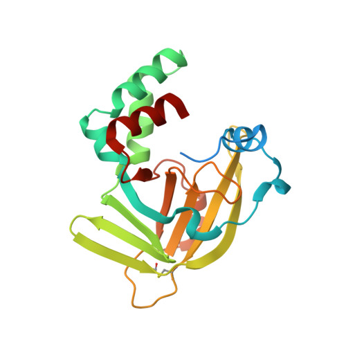 | |
UniProt | |||||
Find proteins for P76344 (Escherichia coli (strain K12)) Explore P76344 Go to UniProtKB: P76344 | |||||
Entity Groups | |||||
| Sequence Clusters | 30% Identity50% Identity70% Identity90% Identity95% Identity100% Identity | ||||
| UniProt Group | P76344 | ||||
Sequence AnnotationsExpand | |||||
| |||||
| Ligands 1 Unique | |||||
|---|---|---|---|---|---|
| ID | Chains | Name / Formula / InChI Key | 2D Diagram | 3D Interactions | |
| ZN Query on ZN | B [auth A] | ZINC ION Zn PTFCDOFLOPIGGS-UHFFFAOYSA-N |  | ||
| Length ( Å ) | Angle ( ˚ ) |
|---|---|
| a = 40.24 | α = 90 |
| b = 65.24 | β = 117.66 |
| c = 41.34 | γ = 90 |
| Software Name | Purpose |
|---|---|
| CBASS | data collection |
| SCALEPACK | data scaling |
| SOLVE | phasing |
| SHARP | phasing |
| CNS | refinement |
RCSB PDB (citation) is hosted by
RCSB PDB is a member of the

