


The crystal structure of conserved hypothetical protein
Min, T., Gorman, J., Shapiro, L.To be published.
Experimental Data Snapshot
wwPDB Validation 3D Report Full Report

Entity ID: 1 | |||||
|---|---|---|---|---|---|
| Molecule | Chains | Sequence Length | Organism | Details | Image |
| conserved hypothetical protein | 139 | Staphylococcus aureus | Mutation(s): 6 | 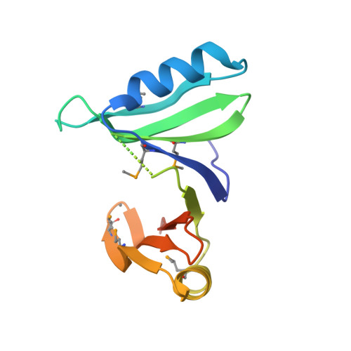 | |
UniProt | |||||
Find proteins for A0A0H3K2T2 (Staphylococcus aureus (strain MW2)) Explore A0A0H3K2T2 Go to UniProtKB: A0A0H3K2T2 | |||||
Entity Groups | |||||
| Sequence Clusters | 30% Identity50% Identity70% Identity90% Identity95% Identity100% Identity | ||||
| UniProt Group | A0A0H3K2T2 | ||||
Sequence AnnotationsExpand | |||||
| |||||
| Modified Residues 1 Unique | |||||
|---|---|---|---|---|---|
| ID | Chains | Type | Formula | 2D Diagram | Parent |
| MSE Query on MSE | A | L-PEPTIDE LINKING | C5 H11 N O2 Se |  | MET |
| Length ( Å ) | Angle ( ˚ ) |
|---|---|
| a = 83.847 | α = 90 |
| b = 83.847 | β = 90 |
| c = 36.52 | γ = 120 |
| Software Name | Purpose |
|---|---|
| REFMAC | refinement |
| DENZO | data reduction |
| SCALEPACK | data scaling |
| SOLVE | phasing |
RCSB PDB (citation) is hosted by
RCSB PDB is a member of the

