1SI8

Crystal structure of E. faecalis catalase


Experimental Data Snapshot

  • Method: X-RAY DIFFRACTION
  • Resolution: 2.30 Å
  • R-Value Free: 0.218 
  • R-Value Work: 0.202 
  • R-Value Observed: 0.202 

wwPDB Validation   3D Report Full Report


Ligand Structure Quality Assessment 


This is version 1.4 of the entry. See complete history


Literature

The three-dimensional structure of catalase from Enterococcus faecalis.

Hakansson, K.O.Brugna, M.Tasse, L.

(2004) Acta Crystallogr D Biol Crystallogr 60: 1374-1380

  • DOI: https://doi.org/10.1107/S0907444904012004
  • Primary Citation of Related Structures:  
    1SI8

  • PubMed Abstract: 

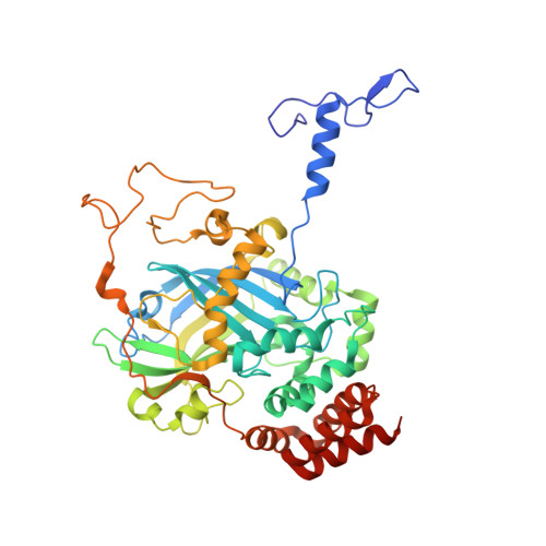
    Enterococcus faecalis haem catalase was crystallized using lithium sulfate at neutral pH. The crystals belong to space group R3, with unit-cell parameters a = b = 236.9, c = 198.1 A. The three-dimensional structure was determined by molecular replacement using a subunit of the Proteus mirabilis catalase structure. It was refined against 2.3 A synchrotron data to a free R factor of 21.8%. Like other catalases, the E. faecalis catalase is a homotetramer with a fold and structure similar to those of its structurally closest relative P. mirabilis. The solvent structure in the active site is identical in the four subunits but differs from that found in other catalases. The structural consequences of the Ramachandran outlier Ser196 are discussed.


  • Organizational Affiliation

    August Krogh Institute, Copenhagen University, Universitetsparken 13, DK-2100 Kbh O, Denmark. kohakansson@aki.ku.dk


Macromolecules
Find similar proteins by:  (by identity cutoff)  |  3D Structure
Entity ID: 1
MoleculeChains Sequence LengthOrganismDetailsImage
Catalase
A, B, C, D
484Enterococcus faecalis V583Mutation(s): 0 
Gene Names: katA
EC: 1.11.1.6
UniProt
Find proteins for Q834P5 (Enterococcus faecalis (strain ATCC 700802 / V583))
Explore Q834P5 
Go to UniProtKB:  Q834P5
Entity Groups  
Sequence Clusters30% Identity50% Identity70% Identity90% Identity95% Identity100% Identity
UniProt GroupQ834P5
Sequence Annotations
Expand
  • Reference Sequence
Small Molecules
Ligands 3 Unique
IDChains Name / Formula / InChI Key2D Diagram3D Interactions
HEM
Query on HEM

Download Ideal Coordinates CCD File 
H [auth A],
K [auth B],
O [auth C],
R [auth D]
PROTOPORPHYRIN IX CONTAINING FE
C34 H32 Fe N4 O4
KABFMIBPWCXCRK-RGGAHWMASA-L
SO4
Query on SO4

Download Ideal Coordinates CCD File 
E [auth A]
F [auth A]
I [auth B]
J [auth B]
L [auth C]
E [auth A],
F [auth A],
I [auth B],
J [auth B],
L [auth C],
M [auth C],
P [auth D],
Q [auth D]
SULFATE ION
O4 S
QAOWNCQODCNURD-UHFFFAOYSA-L
CL
Query on CL

Download Ideal Coordinates CCD File 
G [auth A],
N [auth C]
CHLORIDE ION
Cl
VEXZGXHMUGYJMC-UHFFFAOYSA-M
Experimental Data & Validation

Experimental Data

  • Method: X-RAY DIFFRACTION
  • Resolution: 2.30 Å
  • R-Value Free: 0.218 
  • R-Value Work: 0.202 
  • R-Value Observed: 0.202 
  • Space Group: H 3
Unit Cell:
Length ( Å )Angle ( ˚ )
a = 236.86α = 90
b = 236.86β = 90
c = 198.07γ = 120
Software Package:
Software NamePurpose
MOSFLMdata reduction
SCALAdata scaling
AMoREphasing
CNSrefinement
CCP4data scaling

Structure Validation

View Full Validation Report


Ligand Structure Quality Assessment 


Entry History 

Deposition Data

Revision History  (Full details and data files)

  • Version 1.0: 2004-05-04
    Type: Initial release
  • Version 1.1: 2008-04-29
    Changes: Version format compliance
  • Version 1.2: 2011-07-13
    Changes: Source and taxonomy, Version format compliance
  • Version 1.3: 2018-03-07
    Changes: Data collection
  • Version 1.4: 2023-08-23
    Changes: Data collection, Database references, Derived calculations, Refinement description