1SI1

Crystal Structure of Mannheimia haemolytica Ferric iron-Binding Protein A in an open conformation


Experimental Data Snapshot

  • Method: X-RAY DIFFRACTION
  • Resolution: 1.45 Å
  • R-Value Free: 0.191 
  • R-Value Work: 0.171 
  • R-Value Observed: 0.172 

wwPDB Validation   3D Report Full Report


This is version 1.3 of the entry. See complete history


Literature

Structural basis for iron binding and release by a novel class of periplasmic iron-binding proteins found in gram-negative pathogens.

Shouldice, S.R.Skene, R.J.Dougan, D.R.Snell, G.McRee, D.E.Schryvers, A.B.Tari, L.W.

(2004) J Bacteriol 186: 3903-3910

  • DOI: https://doi.org/10.1128/JB.186.12.3903-3910.2004
  • Primary Citation of Related Structures:  
    1SI0, 1SI1

  • PubMed Abstract: 

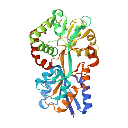
    We have determined the 1.35- and 1.45-A structures, respectively, of closed and open iron-loaded forms of Mannheimia haemolytica ferric ion-binding protein A. M. haemolytica is the causative agent in the economically important and fatal disease of cattle termed shipping fever. The periplasmic iron-binding protein of this gram-negative bacterium, which has homologous counterparts in many other pathogenic species, performs a key role in iron acquisition from mammalian host serum iron transport proteins and is essential for the survival of the pathogen within the host. The ferric (Fe(3+)) ion in the closed structure is bound by a novel asymmetric constellation of four ligands, including a synergistic carbonate anion. The open structure is ligated by three tyrosyl residues and a dynamically disordered solvent-exposed anion. Our results clearly implicate the synergistic anion as the primary mediator of global protein conformation and provide detailed insights into the molecular mechanisms of iron binding and release in the periplasm.


  • Organizational Affiliation

    Department of Microbiology and Infectious Diseases, University of Calgary, Calgary, AB, Canada.


Macromolecules
Find similar proteins by:  (by identity cutoff)  |  3D Structure
Entity ID: 1
MoleculeChains Sequence LengthOrganismDetailsImage
iron binding protein FbpA320Mannheimia haemolyticaMutation(s): 0 
Gene Names: fbpA
UniProt
Find proteins for Q9Z4N6 (Mannheimia haemolytica)
Explore Q9Z4N6 
Go to UniProtKB:  Q9Z4N6
Entity Groups  
Sequence Clusters30% Identity50% Identity70% Identity90% Identity95% Identity100% Identity
UniProt GroupQ9Z4N6
Sequence Annotations
Expand
  • Reference Sequence
Small Molecules
Ligands 1 Unique
IDChains Name / Formula / InChI Key2D Diagram3D Interactions
FE
Query on FE

Download Ideal Coordinates CCD File 
B [auth A]FE (III) ION
Fe
VTLYFUHAOXGGBS-UHFFFAOYSA-N
Experimental Data & Validation

Experimental Data

  • Method: X-RAY DIFFRACTION
  • Resolution: 1.45 Å
  • R-Value Free: 0.191 
  • R-Value Work: 0.171 
  • R-Value Observed: 0.172 
  • Space Group: P 1 21 1
Unit Cell:
Length ( Å )Angle ( ˚ )
a = 46.031α = 90
b = 48.191β = 100.92
c = 77.362γ = 90
Software Package:
Software NamePurpose
REFMACrefinement
HKL-2000data reduction
CCP4data scaling
MOLREPphasing

Structure Validation

View Full Validation Report


Entry History 

Deposition Data

Revision History  (Full details and data files)

  • Version 1.0: 2004-06-08
    Type: Initial release
  • Version 1.1: 2007-10-16
    Changes: Version format compliance
  • Version 1.2: 2011-07-13
    Changes: Version format compliance
  • Version 1.3: 2023-08-23
    Changes: Data collection, Database references, Derived calculations, Refinement description