1S23

Crystal Structure Analysis of the B-DNA Decamer CGCAATTGCG


Experimental Data Snapshot

  • Method: X-RAY DIFFRACTION
  • Resolution: 1.60 Å
  • R-Value Free: 0.282 
  • R-Value Work: 0.210 
  • R-Value Observed: 0.216 

wwPDB Validation   3D Report Full Report


This is version 1.4 of the entry. See complete history


Literature

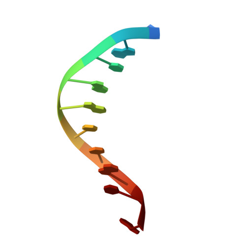
DNA variability in five crystal structures of d(CGCAATTGCG).

Valls, N.Wright, G.Steiner, R.A.Murshudov, G.N.Subirana, J.A.

(2004) Acta Crystallogr D Biol Crystallogr 60: 680-685

  • DOI: https://doi.org/10.1107/S0907444904002896
  • Primary Citation of Related Structures:  
    1S23

  • PubMed Abstract: 

    The deoxyoligonucleotide d(CGCAATTGCG) has previously been crystallized in four different space groups. The crystals diffract to moderate resolution (2.3-2.9 A). Here, a fifth crystal form that diffracts to higher resolution (1.6 A) is presented which was obtained thanks to the use of Co2+ and cryogenic temperatures. The availability of five different crystal structures allows a thorough analysis of the conformational variability of this DNA sequence. It is concluded that the central hexamer sequence CAATTG has a practically constant conformation under all conditions, whilst the terminal base pairs at both ends vary considerably as a result of differing interactions in the crystals. The new crystal structure presented here is stabilized by guanine-Co2+-guanine interactions and the formation of C1+ -G8.C3 triplexes between neighbouring duplexes. As a result of the higher resolution of the crystal structure, a more regular structure was obtained and a clear definition of the spine of hydration was observed which was not visible in the four previous structures.


  • Organizational Affiliation

    Departament d'Enginyeria Química, Universitat Politècnica de Catalunya, Avinguda Diagonal 647, E-08028 Barcelona, Spain.


Macromolecules

Find similar nucleic acids by:  Sequence   |   3D Structure  

Entity ID: 1
MoleculeChains LengthOrganismImage
5'-D(*CP*GP*CP*AP*AP*TP*TP*GP*CP*G)-3'10N/A
Sequence Annotations
Expand
  • Reference Sequence
Small Molecules
Ligands 1 Unique
IDChains Name / Formula / InChI Key2D Diagram3D Interactions
CO
Query on CO

Download Ideal Coordinates CCD File 
B [auth A]COBALT (II) ION
Co
XLJKHNWPARRRJB-UHFFFAOYSA-N
Experimental Data & Validation

Experimental Data

  • Method: X-RAY DIFFRACTION
  • Resolution: 1.60 Å
  • R-Value Free: 0.282 
  • R-Value Work: 0.210 
  • R-Value Observed: 0.216 
  • Space Group: I 21 21 21
Unit Cell:
Length ( Å )Angle ( ˚ )
a = 26.22α = 90
b = 44.359β = 90
c = 52.336γ = 90
Software Package:
Software NamePurpose
REFMACrefinement
DENZOdata reduction
SCALEPACKdata scaling
EPMRphasing

Structure Validation

View Full Validation Report


Entry History 

Deposition Data

Revision History  (Full details and data files)

  • Version 1.0: 2004-04-06
    Type: Initial release
  • Version 1.1: 2008-04-29
    Changes: Version format compliance
  • Version 1.2: 2011-07-13
    Changes: Version format compliance
  • Version 1.3: 2024-02-14
    Changes: Data collection, Database references, Derived calculations
  • Version 1.4: 2024-04-03
    Changes: Refinement description