1R6V

Crystal structure of fervidolysin from Fervidobacterium pennivorans, a keratinolytic enzyme related to subtilisin


Experimental Data Snapshot

  • Method: X-RAY DIFFRACTION
  • Resolution: 1.70 Å
  • R-Value Free: 0.214 
  • R-Value Work: 0.186 
  • R-Value Observed: 0.186 

wwPDB Validation   3D Report Full Report


This is version 1.4 of the entry. See complete history


Literature

Crystal structure of fervidolysin from Fervidobacterium pennivorans, a keratinolytic enzyme related to subtilisin.

Kim, J.S.Kluskens, L.D.de Vos, W.M.Huber, R.van der Oost, J.

(2004) J Mol Biol 335: 787-797

  • DOI: https://doi.org/10.1016/j.jmb.2003.11.006
  • Primary Citation of Related Structures:  
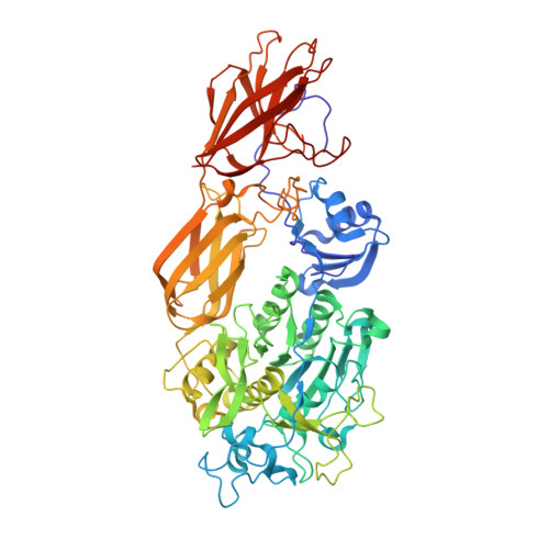
    1R6V

  • PubMed Abstract: 

    Structure-forming fibrous proteins like keratins, gelatins and collagens are degraded only by a few proteases as their tight packing limits access to the potential cleavage sites. To understand the keratin degradation in detail, we describe the first crystal structure of a keratin-degrading enzyme (keratinase), fervidolysin, from Fervidobacterium pennivorans as an immature form with propeptide (PD)-bound. The 1.7A resolution crystal structure shows that the protease is composed of four domains: a catalytic domain (CD), two beta-sandwich domains (SDs), and the PD domain. A structural alignment shows a distant relationship between the PD-CD substructure of fervidolysin and pro-subtilisin E. Tight binding of PD to the remaining part of the protease is mediated by hydrogen bonds along the domain surfaces and around the active cleft, and by the clamps to SD1 and SD2. The crystal structure of this multi-domain protein fervidolysin provides insights into proenzyme activation and the role of non-catalytic domains, suggesting a functional relationship to the fibronectin (FN)-like domains of the human promatrix metalloprotease-2 (proMMP-2) that degrades the fibrous polymeric substrate gelatin.


  • Organizational Affiliation

    Max-Planck-Institut für Biochemie, Abteilung Strukturforschung, Am Klopferspitz 18a, D-82152 Martinsried, Germany. jkim@biochem.mpg.de


Macromolecules
Find similar proteins by:  (by identity cutoff)  |  3D Structure
Entity ID: 1
MoleculeChains Sequence LengthOrganismDetailsImage
subtilisin-like serine protease671Fervidobacterium pennivoransMutation(s): 1 
UniProt
Find proteins for Q93LQ6 (Fervidobacterium pennivorans)
Explore Q93LQ6 
Go to UniProtKB:  Q93LQ6
Entity Groups  
Sequence Clusters30% Identity50% Identity70% Identity90% Identity95% Identity100% Identity
UniProt GroupQ93LQ6
Sequence Annotations
Expand
  • Reference Sequence
Small Molecules
Ligands 1 Unique
IDChains Name / Formula / InChI Key2D Diagram3D Interactions
CA
Query on CA

Download Ideal Coordinates CCD File 
B [auth A]CALCIUM ION
Ca
BHPQYMZQTOCNFJ-UHFFFAOYSA-N
Experimental Data & Validation

Experimental Data

  • Method: X-RAY DIFFRACTION
  • Resolution: 1.70 Å
  • R-Value Free: 0.214 
  • R-Value Work: 0.186 
  • R-Value Observed: 0.186 
  • Space Group: P 1
Unit Cell:
Length ( Å )Angle ( ˚ )
a = 49.558α = 93.9
b = 51.031β = 104.72
c = 79.101γ = 115.43
Software Package:
Software NamePurpose
DENZOdata reduction
SCALEPACKdata scaling
RESOLVEmodel building
CNSrefinement
RESOLVEphasing

Structure Validation

View Full Validation Report


Entry History 

Deposition Data

Revision History  (Full details and data files)

  • Version 1.0: 2004-10-19
    Type: Initial release
  • Version 1.1: 2008-04-29
    Changes: Version format compliance
  • Version 1.2: 2011-07-13
    Changes: Version format compliance
  • Version 1.3: 2021-10-27
    Changes: Database references, Derived calculations
  • Version 1.4: 2024-02-14
    Changes: Data collection