1R4X

Crystal Structure Analys of the Gamma-COPI Appendage domain


Experimental Data Snapshot

  • Method: X-RAY DIFFRACTION
  • Resolution: 1.90 Å
  • R-Value Free: 0.208 
  • R-Value Work: 0.173 
  • R-Value Observed: 0.175 

wwPDB Validation   3D Report Full Report


This is version 1.2 of the entry. See complete history


Literature

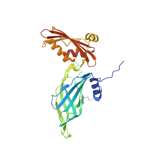
Gamma-COP appendage domain - structure and function

Watson, P.J.Frigerio, G.Collins, B.M.Duden, R.Owen, D.J.

(2004) Traffic 5: 79-88

  • DOI: https://doi.org/10.1111/j.1600-0854.2004.00158.x
  • Primary Citation of Related Structures:  
    1R4X

  • PubMed Abstract: 

    COPI-coated vesicles mediate retrograde transport from the Golgi back to the ER and intra-Golgi transport. The cytosolic precursor of the COPI coat, the heptameric coatomer complex, can be thought of as composed of two subcomplexes. The first consists of the beta-, gamma-, delta- and zeta-COP subunits which are distantly homologous to AP clathrin adaptor subunits. The second consists of the alpha-, beta'- and epsilon-COP subunits. Here, we present the structure of the appendage domain of gamma-COP and show that it has a similar overall fold as the alpha-appendage of AP2. Again, like the alpha-appendage the gamma-COP appendage possesses a single protein/protein interaction site on its platform subdomain. We show that in yeast this site binds to the ARFGAP Glo3p, and in mammalian gamma-COP this site binds to a Glo3p orthologue, ARFGAP2. On the basis of mutations in the yeast homologue of gamma-COP, Sec21p, a second binding site is proposed to exist on the gamma-COP appendage that interacts with the alpha,beta',epsilon COPI subcomplex.


  • Organizational Affiliation

    Cambridge Institute for Medical Research & Department of Clinical Biochemistry, University of Cambridge, Hills Road, Cambridge CB2 2XY, UK.


Macromolecules
Find similar proteins by:  (by identity cutoff)  |  3D Structure
Entity ID: 1
MoleculeChains Sequence LengthOrganismDetailsImage
Coatomer gamma subunit275Homo sapiensMutation(s): 1 
UniProt & NIH Common Fund Data Resources
Find proteins for Q9Y678 (Homo sapiens)
Explore Q9Y678 
Go to UniProtKB:  Q9Y678
PHAROS:  Q9Y678
GTEx:  ENSG00000181789 
Entity Groups  
Sequence Clusters30% Identity50% Identity70% Identity90% Identity95% Identity100% Identity
UniProt GroupQ9Y678
Sequence Annotations
Expand
  • Reference Sequence
Small Molecules
Ligands 1 Unique
IDChains Name / Formula / InChI Key2D Diagram3D Interactions
MG
Query on MG

Download Ideal Coordinates CCD File 
B [auth A]MAGNESIUM ION
Mg
JLVVSXFLKOJNIY-UHFFFAOYSA-N
Modified Residues  1 Unique
IDChains TypeFormula2D DiagramParent
CAS
Query on CAS
A
L-PEPTIDE LINKINGC5 H12 As N O2 SCYS
Experimental Data & Validation

Experimental Data

  • Method: X-RAY DIFFRACTION
  • Resolution: 1.90 Å
  • R-Value Free: 0.208 
  • R-Value Work: 0.173 
  • R-Value Observed: 0.175 
  • Space Group: P 21 21 21
Unit Cell:
Length ( Å )Angle ( ˚ )
a = 48.8α = 90
b = 74.1β = 90
c = 99.2γ = 90
Software Package:
Software NamePurpose
MOSFLMdata reduction
SCALAdata scaling
SHARPphasing
REFMACrefinement
CCP4data scaling

Structure Validation

View Full Validation Report


Entry History 

Deposition Data

Revision History  (Full details and data files)

  • Version 1.0: 2003-10-28
    Type: Initial release
  • Version 1.1: 2008-04-29
    Changes: Version format compliance
  • Version 1.2: 2011-07-13
    Changes: Version format compliance