1QPX

CRYSTAL STRUCTURES OF SELF-CAPPING PAPD CHAPERONE HOMODIMERS


Experimental Data Snapshot

  • Method: X-RAY DIFFRACTION
  • Resolution: 2.40 Å
  • R-Value Free: 0.265 
  • R-Value Work: 0.189 

wwPDB Validation   3D Report Full Report


This is version 1.4 of the entry. See complete history


Literature

Structural basis of chaperone self-capping in P pilus biogenesis.

Hung, D.L.Pinkner, J.S.Knight, S.D.Hultgren, S.J.

(1999) Proc Natl Acad Sci U S A 96: 8178-8183

  • DOI: https://doi.org/10.1073/pnas.96.14.8178
  • Primary Citation of Related Structures:  
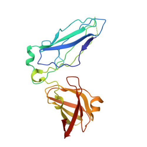
    1QPP, 1QPX

  • PubMed Abstract: 

    PapD is an immunoglobulin-like chaperone that mediates the assembly of P pili in uropathogenic strains of Escherichia coli. It binds and caps interactive surfaces on pilus subunits to prevent their premature associations in the periplasm. We elucidated the structural basis of a mechanism whereby PapD also interacts with itself, capping its own subunit binding surface. Crystal structures of dimeric forms of PapD revealed that this self-capping mechanism involves a rearrangement and ordering of the C2-D2 and F1-G1 loops upon dimerization which might ensure that a stable dimer is not formed in solution in spite of a relatively large dimer interface. An analysis of site directed mutations revealed that chaperone dimerization requires the same surface that is otherwise used to bind subunits.


  • Organizational Affiliation

    Department of Molecular Microbiology, Box 8230, Washington University School of Medicine, 660 South Euclid Avenue, St. Louis, MO 63110, USA.


Macromolecules
Find similar proteins by:  (by identity cutoff)  |  3D Structure
Entity ID: 1
MoleculeChains Sequence LengthOrganismDetailsImage
PAPD CHAPERONE
A, B
218Escherichia coliMutation(s): 1 
UniProt
Find proteins for P15319 (Escherichia coli)
Explore P15319 
Go to UniProtKB:  P15319
Entity Groups  
Sequence Clusters30% Identity50% Identity70% Identity90% Identity95% Identity100% Identity
UniProt GroupP15319
Sequence Annotations
Expand
  • Reference Sequence
Experimental Data & Validation

Experimental Data

  • Method: X-RAY DIFFRACTION
  • Resolution: 2.40 Å
  • R-Value Free: 0.265 
  • R-Value Work: 0.189 
  • Space Group: P 21 21 21
Unit Cell:
Length ( Å )Angle ( ˚ )
a = 53.065α = 90
b = 75.442β = 90
c = 115.189γ = 90
Software Package:
Software NamePurpose
DENZOdata reduction
SCALEPACKdata scaling
AMoREphasing
REFMACrefinement

Structure Validation

View Full Validation Report


Entry History 

Deposition Data

Revision History  (Full details and data files)

  • Version 1.0: 1999-07-07
    Type: Initial release
  • Version 1.1: 2008-04-27
    Changes: Version format compliance
  • Version 1.2: 2011-07-13
    Changes: Version format compliance
  • Version 1.3: 2018-03-14
    Changes: Database references
  • Version 1.4: 2022-12-21
    Changes: Database references