The High Resolution Crystal Structure of Recombinant Crithidia Fasciculata Tryparedoxin-I
Alphey, M.S., Leonard, G.A., Gourley, D.G., Tetaud, E., Fairlamb, A.H., Hunter, W.N.(1999) J Biol Chem 274: 25613
- PubMed: 10464297
- DOI: https://doi.org/10.1074/jbc.274.36.25613
- Primary Citation of Related Structures:
1QK8 - PubMed Abstract:
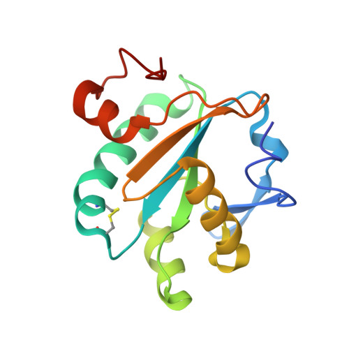
Tryparedoxin-I is a recently discovered thiol-disulfide oxidoreductase involved in the regulation of oxidative stress in parasitic trypanosomatids. The crystal structure of recombinant Crithidia fasciculata tryparedoxin-I in the oxidized state has been determined using multi-wavelength anomalous dispersion methods applied to a selenomethionyl derivative. The model comprises residues 3 to 145 with 236 water molecules and has been refined using all data between a 19- and 1.4-A resolution to an R-factor and R-free of 19.1 and 22.3%, respectively. Despite sharing only about 20% sequence identity, tryparedoxin-I presents a five-stranded twisted beta-sheet and two elements of helical structure in the same type of fold as displayed by thioredoxin, the archetypal thiol-disulfide oxidoreductase. However, the relationship of secondary structure with the linear amino acid sequences is different for each protein, producing a distinctive topology. The beta-sheet core is extended in the trypanosomatid protein with an N-terminal beta-hairpin. There are also differences in the content and orientation of helical elements of secondary structure positioned at the surface of the proteins, which leads to different shapes and charge distributions between human thioredoxin and tryparedoxin-I. A right-handed redox-active disulfide is formed between Cys-40 and Cys-43 at the N-terminal region of a distorted alpha-helix (alpha1). Cys-40 is solvent-accessible, and Cys-43 is positioned in a hydrophilic cavity. Three C-H...O hydrogen bonds donated from two proline residues serve to stabilize the disulfide-carrying helix and support the correct alignment of active site residues. The accurate model for tryparedoxin-I allows for comparisons with the family of thiol-disulfide oxidoreductases and provides a template for the discovery or design of selective inhibitors of hydroperoxide metabolism in trypanosomes. Such inhibitors are sought as potential therapies against a range of human pathogens.
Organizational Affiliation:
Department of Biochemistry, The Wellcome Trust Building, University of Dundee, Dundee, DD1 5EH, Scotland, United Kingdom.