1QJM

Crystal Structure of a Complex of Lactoferrin with a Lanthanide Ion (SM3+) at 3.4 Angstrom Resolution


Experimental Data Snapshot

  • Method: X-RAY DIFFRACTION
  • Resolution: 3.40 Å
  • R-Value Free: 0.316 
  • R-Value Work: 0.219 
  • R-Value Observed: 0.219 

wwPDB Validation   3D Report Full Report


This is version 1.4 of the entry. See complete history


Literature

Lactoferrin-Metal Interactions: First Crystal Structure of a Complex of Lactoferrin with a Lanthanide Ion (Sm3+) at 3.4 Angstrom Resolution

Sharma, A.K.Singh, T.P.

(1999) Acta Crystallogr D Biol Crystallogr 55: 1799

  • DOI: https://doi.org/10.1107/s0907444999009865
  • Primary Citation of Related Structures:  
    1QJM

  • PubMed Abstract: 

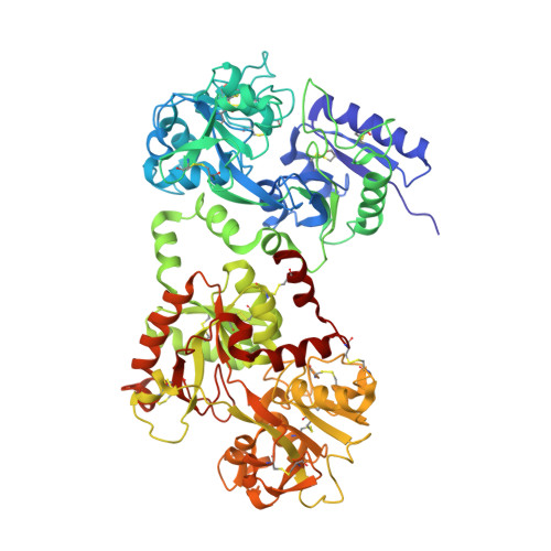
    Lactoferrin is an important member of the transferrin family. A characteristic property of transferrins is their ability to bind very tightly (K(app) approximately/= 10(20)) but reversibly two Fe(3+) ions. The structural consequences of binding a metal other than Fe(3+) have been examined by crystallographic analysis at 3.4 A resolution of mare samarium-lactoferrin (Sm(2)Lf). The structure was refined to an R factor of 0.219 for 8776 reflections in the resolution range 17.0-3.4 A. The samarium geometry (distorted octahedral coordination) is similar in both lobes. However, the anion interactions are quite different in the two lobes. In the N lobe, the anion is able to form only two hydrogen bonds instead of the four observed in the C lobe of Sm(2)Lf and the six observed in Fe(2)Lf. This is because Arg121, Thr117 and Gly124 have moved away from the anion as a consequence of the binding of the Sm(3+) ion. The protein ligands in the binding cleft of Sm(2)Lf show large displacements, but the overall protein structure remains the same. The binding of Sm(3+) by lactoferrin shows that the protein is capable of sequestering ions of different sizes and charges, though with reduced affinity. This conclusion should be true of other transferrins also.


  • Organizational Affiliation

    Department of Biophysics, All India Institute of Medical Sciences, New Delhi 110 029, India.


Macromolecules
Find similar proteins by:  (by identity cutoff)  |  3D Structure
Entity ID: 1
MoleculeChains Sequence LengthOrganismDetailsImage
LACTOFERRIN689Equus caballusMutation(s): 0 
UniProt
Find proteins for O77811 (Equus caballus)
Explore O77811 
Go to UniProtKB:  O77811
Entity Groups  
Sequence Clusters30% Identity50% Identity70% Identity90% Identity95% Identity100% Identity
UniProt GroupO77811
Sequence Annotations
Expand
  • Reference Sequence
Experimental Data & Validation

Experimental Data

  • Method: X-RAY DIFFRACTION
  • Resolution: 3.40 Å
  • R-Value Free: 0.316 
  • R-Value Work: 0.219 
  • R-Value Observed: 0.219 
  • Space Group: P 21 21 21
Unit Cell:
Length ( Å )Angle ( ˚ )
a = 80.492α = 90
b = 103.191β = 90
c = 113.16γ = 90
Software Package:
Software NamePurpose
X-PLORrefinement
DENZOdata reduction
SCALEPACKdata scaling
X-PLORphasing

Structure Validation

View Full Validation Report


Entry History 

Deposition Data

Revision History  (Full details and data files)

  • Version 1.0: 1999-08-26
    Type: Initial release
  • Version 1.1: 2016-12-28
    Changes: Database references, Derived calculations, Other, Structure summary, Version format compliance
  • Version 1.2: 2017-07-05
    Changes: Data collection
  • Version 1.3: 2019-04-03
    Changes: Data collection, Experimental preparation
  • Version 1.4: 2023-12-13
    Changes: Data collection, Database references, Derived calculations, Refinement description