A structural view of evolutionary divergence.
Spiller, B., Gershenson, A., Arnold, F.H., Stevens, R.C.(1999) Proc Natl Acad Sci U S A 96: 12305-12310
- PubMed: 10535917
- DOI: https://doi.org/10.1073/pnas.96.22.12305
- Primary Citation of Related Structures:
1C7I, 1C7J, 1QE3 - PubMed Abstract:
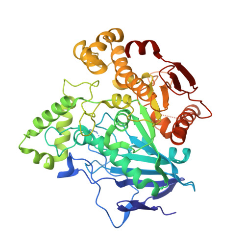
Two directed evolution experiments on p-nitrobenzyl esterase yielded one enzyme with a 100-fold increased activity in aqueous-organic solvents and another with a 17 degrees C increase in thermostability. Structures of the wild type and its organophilic and thermophilic counterparts are presented at resolutions of 1.5 A, 1.6 A, and 2.0 A, respectively. These structures identify groups of interacting mutations and demonstrate how directed evolution can traverse complex fitness landscapes. Early-generation mutations stabilize flexible loops not visible in the wild-type structure and set the stage for further beneficial mutations in later generations. The mutations exert their influence on the esterase structure over large distances, in a manner that would be difficult to predict. The loops with the largest structural changes generally are not the sites of mutations. Similarly, none of the seven amino acid substitutions in the organophile are in the active site, even though the enzyme experiences significant changes in the organization of this site. In addition to reduction of surface loop flexibility, thermostability in the evolved esterase results from altered core packing, helix stabilization, and the acquisition of surface salt bridges, in agreement with other comparative studies of mesophilic and thermophilic enzymes. Crystallographic analysis of the wild type and its evolved counterparts reveals networks of mutations that collectively reorganize the active site. Interestingly, the changes that led to diversity within the alpha/beta hydrolase enzyme family and the reorganization seen in this study result from main-chain movements.
Organizational Affiliation:
Department of Molecular Biology, University of California, Berkeley, CA 92037, USA.