The crystal structure of the formiminotransferase domain of formiminotransferase-cyclodeaminase: implications for substrate channeling in a bifunctional enzyme.
Kohls, D., Sulea, T., Purisima, E.O., MacKenzie, R.E., Vrielink, A.(2000) Structure 8: 35-46
- PubMed: 10673422
- DOI: https://doi.org/10.1016/s0969-2126(00)00078-2
- Primary Citation of Related Structures:
1QD1 - PubMed Abstract:
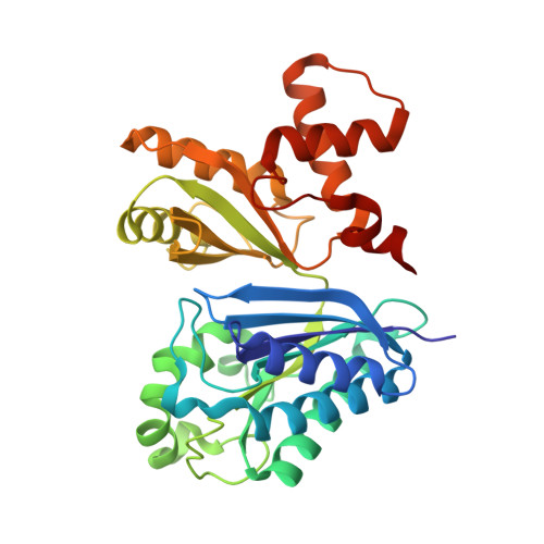
The bifunctional enzyme formiminotransferase-cyclodeaminase (FTCD) contains two active sites at different positions on the protein structure. The enzyme binds a gamma-linked polyglutamylated form of the tetrahydrofolate substrate and channels the product of the transferase reaction from the transferase active site to the cyclodeaminase active site. Structural studies of this bifunctional enzyme and its monofunctional domains will provide insight into the mechanism of substrate channeling and the two catalytic reactions. The crystal structure of the formiminotransferase (FT) domain of FTCD has been determined in the presence of a product analog, folinic acid. The overall structure shows that the FT domain comprises two subdomains that adopt a novel alpha/beta fold. Inspection of the folinic acid binding site reveals an electrostatic tunnel traversing the width of the molecule. The distribution of charged residues in the tunnel provides insight into the possible mode of substrate binding and channeling. The electron density reveals that the non-natural stereoisomer, (6R)-folinic acid, binds to the protein; this observation suggests a mechanism for product release. In addition, a single molecule of glycerol is bound to the enzyme and indicates a putative binding site for formiminoglutamate. The structure of the FT domain in the presence of folinic acid reveals a possible novel mechanism for substrate channeling. The position of the folinic acid and a bound glycerol molecule near to the sidechain of His82 suggests that this residue may act as the catalytic base required for the formiminotransferase mechanism.
Organizational Affiliation:
Biochemistry Department, McIntyre Medical Sciences Building, Montréal Joint Center for Structural Biology, McGill University, Montréal, H3G 1Y6, Canada.