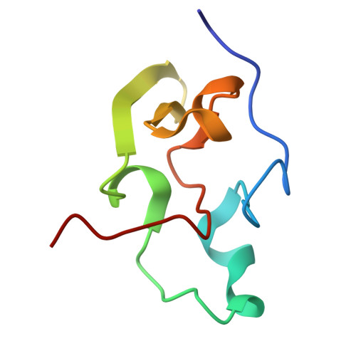
The three-dimensional structure in solution of the paramagnetic high-potential iron-sulfur protein I from Ectothiorhodospira halophila through nuclear magnetic resonance.
Banci, L., Bertini, I., Eltis, L.D., Felli, I.C., Kastrau, D.H., Luchinat, C., Piccioli, M., Pierattelli, R., Smith, M.(1994) Eur J Biochem 225: 715-725
- PubMed: 7957187
- DOI: https://doi.org/10.1111/j.1432-1033.1994.00715.x
- Primary Citation of Related Structures:
1PIH, 1PIJ - PubMed Abstract:
The three-dimensional structure in solution of reduced recombinant high-potential iron-sulfur protein iso-I from Ectothiorhodospira halophila was determined using 948 relevant interproton NOEs out of the 1246 observed NOEs. The determination was accomplished using the XEASY program for spectral analysis and the distance geometry (DG) program DIANA for generation of the structure as described by Wüthrich [Wüthrich, K. (1989) Acc. Chem. Res. 22, 36-44]. The FeS cluster was simulated using an amino acid residue constructed for the present work from a cysteinyl residue with an iron and a sulfur atom attached to the terminal thiol. The family of structures obtained from distance geometry were subjected to energy minimization and molecular dynamics simulations using previously defined force field parameters. The quality of these structures at each stage of the refinement process is discussed with respect to the dihedral angle order parameter and the root-mean-square deviation of the atomic coordinates. The latter values for the backbone atoms vary from 67 pm for the distance-geometry structures to 60 pm for the energy-minimized structures to 51 pm for the structures subjected to restrained molecular dynamics. Finally, the structure in best agreement with the NOE constraints has been further treated with extensive restrained molecular dynamics in water. The solution structure is well defined and is very similar to the available X-ray structure. We do not know of any previous determination of the structure of a paramagnetic protein in solution by NMR. The effect of paramagnetism on the quality of the structure determination is discussed.
Organizational Affiliation:
Department of Chemistry, University of Florence, Italy.