Solution structure and dynamics of PEC-60, a protein of the Kazal type inhibitor family, determined by nuclear magnetic resonance spectroscopy.
Liepinsh, E., Berndt, K.D., Sillard, R., Mutt, V., Otting, G.(1994) J Mol Biol 239: 137-153
- PubMed: 8196042
- DOI: https://doi.org/10.1006/jmbi.1994.1356
- Primary Citation of Related Structures:
1PCE - PubMed Abstract:
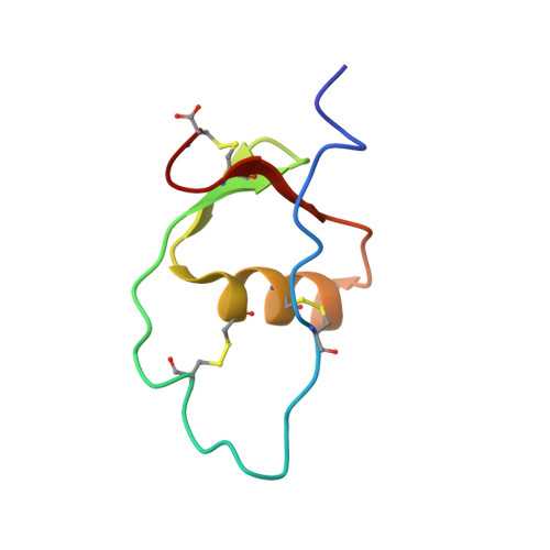
The three-dimensional solution structure of porcine PEC-60, a 60 amino acid residue protein of the Kazal type family of proteinase inhibitors, was determined by nuclear magnetic resonance (NMR) spectroscopy. The structure determination is based on nearly complete 1H, 13C and 15N resonance assignments including stereospecific 1H resonance assignments for 40 pairs of methylene protons and isopropyl methyl groups. The stereospecific resonance assignments of the beta-protons were supported by heteronuclear long-range correlation experiments recorded at natural 13C and 15N isotopic abundances. A group of 20 conformers were calculated using the experimentally derived NMR constraints with the program DIANA, and energy-minimized in a 4 A water shell using the program OPAL. The average of the root-mean-square deviations relative to the mean structure of the 20 conformers selected to represent the solution structure of PEC-60 is 0.55 A for the backbone atoms of residues 6 to 10 and 24 to 60. Disordered conformations are observed for the amino-terminal pentapeptide and the polypeptide segment containing residues 11 to 23. The NMR structure confirms the structural similarity of PEC-60 to the Kazal type family of proteinase inhibitors which had been previously suggested on the basis of amino acid homology. The well-defined part of PEC-60 contains a short three-stranded anti-parallel beta-sheet involving the residues 27 to 29, 33 to 35 and 53 to 56 with a beta-bulge at residue 55, a type I turn comprising residues 29 to 32, and an alpha-helix involving the residues 37 to 48. T1(13C) relaxation measurements of the alpha-carbons and linewidth measurements of the amide proton signals indicate substantially increased mobility on the pico- to nanosecond time-scale for the amino-terminal pentapeptide as well as within the loop comprising residues 11 to 23. The structure of PEC-60 is compared to the X-ray crystal structures of homologous Kazal type proteinase inhibitors and the dynamic properties of PEC-60 are discussed with respect to the observed lack of any substantial trypsin inhibiting activity.
Organizational Affiliation:
Department of Medical Biochemistry and Biophysics, Karolinska Institute, Stockholm, Sweden.