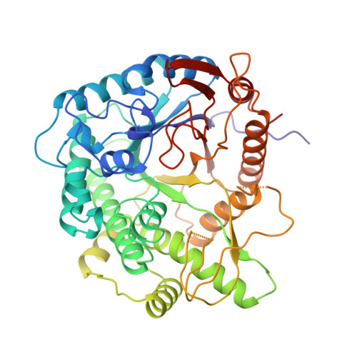
The three-dimensional structure of 6-phospho-beta-galactosidase from Lactococcus lactis.
Wiesmann, C., Beste, G., Hengstenberg, W., Schulz, G.E.(1995) Structure 3: 961-968
- PubMed: 8535789
- DOI: https://doi.org/10.1016/s0969-2126(01)00230-1
- Primary Citation of Related Structures:
1PBG - PubMed Abstract:
The enzyme 6-phospho-beta-galactosidase hydrolyzes phospholactose, the product of a phosphor-enolpyruvate-dependent phosphotransferase system. It belongs to glycosidase family 1 and no structure has yet been published for a member of this family. The crystal structure of 6-phospho-beta-galactosidase was determined at 2.3 A resolution by multiple isomorphous replacement, using the wild-type enzyme and a designed cysteine mutant. A second crystal form, found with the mutant enzyme, was solved by molecular replacement, yielding the conformation of two chain loops that are invisible in the first crystal form. The active center, located through catalytic residues identified in previous studies, cannot be accessed by the substrate if the two loops are in their defined conformation. The enzyme contains a (beta alpha)8 barrel and the relationship of its chain fold to that of other glycosidases has been quantified. As a side issue, we observed that a cysteine point mutant designed for X-ray analysis crystallized mainly as a symmetric dimer around an intermolecular disulfide bridge formed by the newly introduced cysteine. The presented analysis provides a basis on which to model all other family 1 members and thereby will help in elucidating the catalytic mechanisms of these sequence-related enzymes. Moreover, this enzyme belongs to a superfamily of glycosidases sharing a (beta alpha)8 barrel with catalytic glutamates/aspartates at the ends of the fourth and the seventh strands of the beta barrel.
Organizational Affiliation:
Institut für Organische Chemie und Biochemie, Albert-Ludwigs-Universität, Freiburg, Germany.