1ONS

Crystal structure of Escherichia coli heat shock protein YedU


Experimental Data Snapshot

  • Method: X-RAY DIFFRACTION
  • Resolution: 2.20 Å
  • R-Value Free: 0.241 
  • R-Value Work: 0.204 
  • R-Value Observed: 0.220 

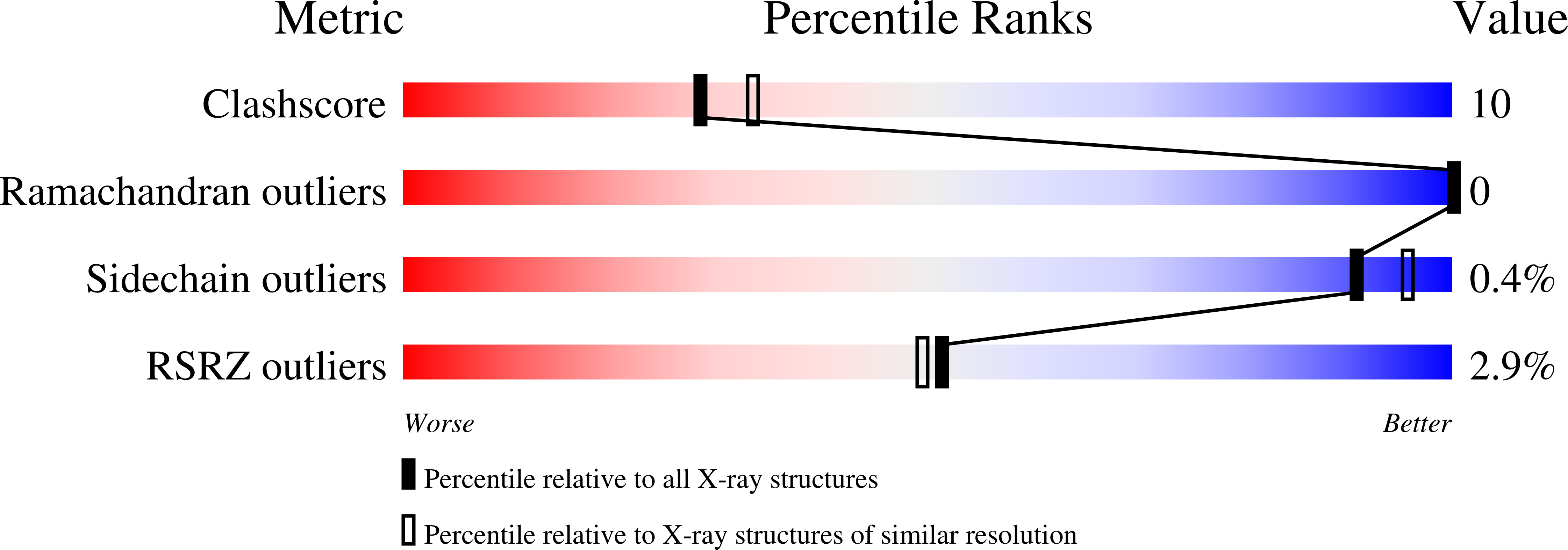
wwPDB Validation   3D Report Full Report


This is version 1.3 of the entry. See complete history


Literature

The crystal structure of Escherichia coli heat shock protein YedU reveals three potential catalytic active sites

Zhao, Y.Liu, D.Kaluarachchi, W.D.Bellamy, H.D.White, M.A.Fox, R.O.

(2003) Protein Sci 12: 2303-2311

  • DOI: https://doi.org/10.1110/ps.03121403
  • Primary Citation of Related Structures:  
    1ONS

  • PubMed Abstract: 

    The mRNA of Escherichia coli yedU gene is induced 31-fold upon heat shock. The 31-kD YedU protein, also calls Hsp31, is highly conserved in several human pathogens and has chaperone activity. We solved the crystal structure of YedU at 2.2 A resolution. YedU monomer has an alpha/beta/alpha sandwich domain and a small alpha/beta domain. YedU is a dimer in solution, and its crystal structure indicates that a significant amount of surface area is buried upon dimerization. There is an extended hydrophobic patch that crosses the dimer interface on the surface of the protein. This hydrophobic patch is likely the substrate-binding site responsible for the chaperone activity. The structure also reveals a potential protease-like catalytic triad composed of Cys184, His185, and Asp213, although no enzymatic activity could be identified. YedU coordinates a metal ion using His85, His122, and Glu90. This 2-His-1-carboxylate motif is present in carboxypeptidase A (a zinc enzyme), and a number of dioxygenases and hydroxylases that utilize iron as a cofactor, suggesting another potential function for YedU.


  • Organizational Affiliation

    Department of Physiology and Biophysics, and The Graduate Program in Cellular Physiology and Molecular Biophysics, The University of Texas Medical Branch at Galveston, Galveston, Texas 77555, USA.


Macromolecules
Find similar proteins by:  (by identity cutoff)  |  3D Structure
Entity ID: 1
MoleculeChains Sequence LengthOrganismDetailsImage
Chaperone protein hchA282Escherichia coliMutation(s): 0 
Gene Names: HCHA OR B1967
UniProt
Find proteins for P31658 (Escherichia coli (strain K12))
Explore P31658 
Go to UniProtKB:  P31658
Entity Groups  
Sequence Clusters30% Identity50% Identity70% Identity90% Identity95% Identity100% Identity
UniProt GroupP31658
Sequence Annotations
Expand
  • Reference Sequence
Small Molecules
Ligands 1 Unique
IDChains Name / Formula / InChI Key2D Diagram3D Interactions
ZN
Query on ZN

Download Ideal Coordinates CCD File 
B [auth A]ZINC ION
Zn
PTFCDOFLOPIGGS-UHFFFAOYSA-N
Experimental Data & Validation

Experimental Data

  • Method: X-RAY DIFFRACTION
  • Resolution: 2.20 Å
  • R-Value Free: 0.241 
  • R-Value Work: 0.204 
  • R-Value Observed: 0.220 
  • Space Group: P 61 2 2
Unit Cell:
Length ( Å )Angle ( ˚ )
a = 52.98α = 90
b = 52.98β = 90
c = 347.4γ = 120
Software Package:
Software NamePurpose
DENZOdata reduction
SCALEPACKdata scaling
XFITdata reduction
CNSrefinement

Structure Validation

View Full Validation Report


Entry History 

Deposition Data

Revision History  (Full details and data files)

  • Version 1.0: 2004-01-20
    Type: Initial release
  • Version 1.1: 2008-04-29
    Changes: Version format compliance
  • Version 1.2: 2011-07-13
    Changes: Derived calculations, Version format compliance
  • Version 1.3: 2024-02-14
    Changes: Data collection, Database references, Derived calculations