1OJI

Anatomy of glycosynthesis: Structure and kinetics of the Humicola insolens Cel7B E197A and E197S glycosynthase mutants


Experimental Data Snapshot

  • Method: X-RAY DIFFRACTION
  • Resolution: 2.15 Å
  • R-Value Free: 0.226 
  • R-Value Work: 0.192 
  • R-Value Observed: 0.194 

wwPDB Validation   3D Report Full Report


Ligand Structure Quality Assessment 


This is version 2.2 of the entry. See complete history


Literature

Anatomy of Glycosynthesis: Structure and Kinetics of the Humicola Insolens Cel7B E197A and E197S Glycosynthase Mutants

Ducros, V.M.-A.Tarling, C.A.Zechel, D.L.Brzozowski, A.M.Frandsen, T.P.Von Ossowski, I.Schulein, M.Withers, S.G.Davies, G.J.

(2003) Chem Biol 10: 619

  • DOI: https://doi.org/10.1016/s1074-5521(03)00143-1
  • Primary Citation of Related Structures:  
    1OJI, 1OJJ, 1OJK

  • PubMed Abstract: 

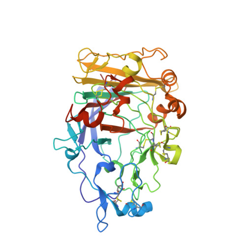
    The formation of glycoconjugates and oligosaccharides remains one of the most challenging chemical syntheses. Chemo-enzymatic routes using retaining glycosidases have been successfully harnessed but require tight kinetic or thermodynamic control. "Glycosynthases," specifically engineered glycosidases that catalyze the formation of glycosidic bonds from glycosyl donor and acceptor alcohol, are an emerging range of synthetic tools in which catalytic nucleophile mutants are harnessed together with glycosyl fluoride donors to generate powerful and versatile catalysts. Here we present the structural and kinetic dissection of the Humicola insolens Cel7B glycosynthases in which the nucleophile of the wild-type enzyme is mutated to alanine and serine (E197A and E197S). 3-D structures reveal the acceptor and donor subsites and the basis for substrate inhibition. Kinetic analysis shows that the E197S mutant is considerably more active than the corresponding alanine mutant due to a 40-fold increase in k(cat).


  • Organizational Affiliation

    Structural Biology Laboratory, Department of Chemistry, The University of York, Heslington, York YO10 5YW, United Kingdom.


Macromolecules
Find similar proteins by:  (by identity cutoff)  |  3D Structure
Entity ID: 1
MoleculeChains Sequence LengthOrganismDetailsImage
ENDOGLUCANASE I402Humicola insolensMutation(s): 1 
EC: 3.2.1.4
UniProt
Find proteins for P56680 (Humicola insolens)
Explore P56680 
Go to UniProtKB:  P56680
Entity Groups  
Sequence Clusters30% Identity50% Identity70% Identity90% Identity95% Identity100% Identity
UniProt GroupP56680
Sequence Annotations
Expand
  • Reference Sequence
Small Molecules
Ligands 2 Unique
IDChains Name / Formula / InChI Key2D Diagram3D Interactions
NAG
Query on NAG

Download Ideal Coordinates CCD File 
B [auth A]2-acetamido-2-deoxy-beta-D-glucopyranose
C8 H15 N O6
OVRNDRQMDRJTHS-FMDGEEDCSA-N
GOL
Query on GOL

Download Ideal Coordinates CCD File 
C [auth A]GLYCEROL
C3 H8 O3
PEDCQBHIVMGVHV-UHFFFAOYSA-N
Modified Residues  1 Unique
IDChains TypeFormula2D DiagramParent
PCA
Query on PCA
A
L-PEPTIDE LINKINGC5 H7 N O3GLN
Experimental Data & Validation

Experimental Data

  • Method: X-RAY DIFFRACTION
  • Resolution: 2.15 Å
  • R-Value Free: 0.226 
  • R-Value Work: 0.192 
  • R-Value Observed: 0.194 
  • Space Group: P 65
Unit Cell:
Length ( Å )Angle ( ˚ )
a = 122.312α = 90
b = 122.312β = 90
c = 82.616γ = 120
Software Package:
Software NamePurpose
REFMACrefinement
DENZOdata reduction
SCALEPACKdata scaling
AMoREphasing

Structure Validation

View Full Validation Report


Ligand Structure Quality Assessment 


Entry History 

Deposition Data

Revision History  (Full details and data files)

  • Version 1.0: 2004-01-07
    Type: Initial release
  • Version 1.1: 2011-05-08
    Changes: Version format compliance
  • Version 1.2: 2011-07-13
    Changes: Version format compliance
  • Version 1.3: 2019-05-08
    Changes: Data collection, Derived calculations, Experimental preparation, Other
  • Version 2.0: 2020-03-11
    Changes: Data collection, Other, Polymer sequence
  • Version 2.1: 2020-07-29
    Type: Remediation
    Reason: Carbohydrate remediation
    Changes: Data collection, Derived calculations, Structure summary
  • Version 2.2: 2023-12-13
    Changes: Data collection, Database references, Refinement description, Structure summary