1ODG

Very-short-patch DNA repair endonuclease bound to its reaction product site


Experimental Data Snapshot

  • Method: X-RAY DIFFRACTION
  • Resolution: 2.80 Å
  • R-Value Free: 0.356 
  • R-Value Work: 0.287 
  • R-Value Observed: 0.287 

wwPDB Validation   3D Report Full Report


This is version 1.3 of the entry. See complete history


Literature

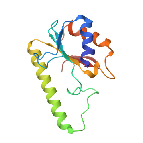
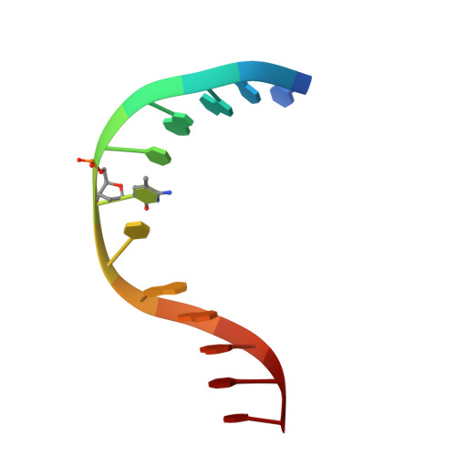
Crystal Structure of the Escherichia Coli Dcm Very-Short-Patch DNA Repair Endonuclease Bound to its Reaction Product-Site in a DNA Superhelix

Bunting, K.A.Roe, S.M.Headley, A.Brown, T.Savva, R.Pearl, L.H.

(2003) Nucleic Acids Res 31: 1633

  • DOI: https://doi.org/10.1093/nar/gkg273
  • Primary Citation of Related Structures:  
    1ODG

  • PubMed Abstract: 

    Very-short-patch repair (Vsr) enzymes occur in a variety of bacteria, where they initiate nucleotide excision repair of G:T mismatches arising by deamination of 5-methyl-cytosines in specific regulatory sequences. We have now determined the structure of the archetypal dcm-Vsr endonuclease from Escherichia coli bound to the cleaved authentic hemi-deaminated/hemi-methylated dcm sequence 5'-C-OH-3' 5'-p-T-p-A-p-G-p-G-3'/3'-G-p-G-p-T-p(Me5)C-p-C formed by self-assembly of a 12mer oligonucleotide into a continuous nicked DNA superhelix. The structure reveals the presence of a Hoogsteen base pair within the deaminated recognition sequence and the substantial distortions of the DNA that accompany Vsr binding to product sites.


  • Organizational Affiliation

    Section of Structural Biology, Institute of Cancer Research, Chester Beatty Laboratories, 237 Fulham Road, London SW3 6JB, UK.


Macromolecules

Find similar proteins by:  (by identity cutoff)  |  3D Structure
Entity ID: 1
MoleculeChains Sequence LengthOrganismDetailsImage
DNA MISMATCH ENDONUCLEASE134Escherichia coliMutation(s): 0 
UniProt
Find proteins for P09184 (Escherichia coli (strain K12))
Explore P09184 
Go to UniProtKB:  P09184
Entity Groups  
Sequence Clusters30% Identity50% Identity70% Identity90% Identity95% Identity100% Identity
UniProt GroupP09184
Sequence Annotations
Expand
  • Reference Sequence

Find similar nucleic acids by:  Sequence   |   3D Structure  

Entity ID: 2
MoleculeChains LengthOrganismImage
5'-D(*TP*AP*GP*GP*CP*5CM*TP*GP*GP*AP*TP*CP)-3'B [auth F],
C [auth W]
12Escherichia coli
Sequence Annotations
Expand
  • Reference Sequence
Small Molecules
Ligands 1 Unique
IDChains Name / Formula / InChI Key2D Diagram3D Interactions
ZN
Query on ZN

Download Ideal Coordinates CCD File 
D [auth A]ZINC ION
Zn
PTFCDOFLOPIGGS-UHFFFAOYSA-N
Experimental Data & Validation

Experimental Data

  • Method: X-RAY DIFFRACTION
  • Resolution: 2.80 Å
  • R-Value Free: 0.356 
  • R-Value Work: 0.287 
  • R-Value Observed: 0.287 
  • Space Group: P 61
Unit Cell:
Length ( Å )Angle ( ˚ )
a = 102.805α = 90
b = 102.805β = 90
c = 64.29γ = 120
Software Package:
Software NamePurpose
CNSrefinement
MOSFLMdata reduction
SCALAdata scaling
AMoREphasing

Structure Validation

View Full Validation Report


Entry History 

Deposition Data

Revision History  (Full details and data files)

  • Version 1.0: 2003-03-13
    Type: Initial release
  • Version 1.1: 2011-05-08
    Changes: Version format compliance
  • Version 1.2: 2011-07-13
    Changes: Version format compliance
  • Version 1.3: 2023-12-13
    Changes: Data collection, Database references, Derived calculations, Other, Refinement description