1NZE

Crystal structure of PsbQ polypeptide of photosystem II from higher plants


Experimental Data Snapshot

  • Method: X-RAY DIFFRACTION
  • Resolution: 1.95 Å
  • R-Value Free: 0.268 
  • R-Value Work: 0.227 
  • R-Value Observed: 0.227 

wwPDB Validation   3D Report Full Report


This is version 1.3 of the entry. See complete history


Literature

Crystal structure of the PsbQ protein of photosystem II from higher plants

Calderone, V.Trabucco, M.Vujicic, A.Battistutta, R.Giacometti, G.M.Andreucci, F.Barbato, R.Zanotti, G.

(2003) EMBO Rep 4: 900-905

  • DOI: https://doi.org/10.1038/sj.embor.embor923
  • Primary Citation of Related Structures:  
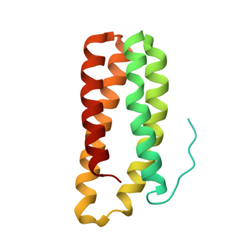
    1NZE

  • PubMed Abstract: 

    The smallest extrinsic polypeptide of the water-oxidizing complex (PsbQ) was extracted and purified from spinach (Spinacia oleracea) photosystem II (PSII) membranes. It was then crystallized in the presence of Zn(2+) and its structure was determined by X-ray diffraction at 1.95-A resolution using the multi-wavelength anomalous diffraction method, with the zinc as the anomalous scatterer. The crystal structure shows that the core of the protein is a four-helix bundle, whereas the amino-terminal portion, which possibly interacts with the photosystem core, is not visible in the crystal. The distribution of positive and negative charges on the protein surface might explain the ability of PsbQ to increase the binding of Cl(-) and Ca(2+) and make them available to PSII.


  • Organizational Affiliation

    Dipartimento di Chimica Organica e Centro Biopolimeri del CNR, Università di Padova, Via Marzolo 1, 35131 Padua, Italy.


Macromolecules
Find similar proteins by:  (by identity cutoff)  |  3D Structure
Entity ID: 1
MoleculeChains Sequence LengthOrganismDetailsImage
Oxygen-evolving enhancer protein 3149Spinacia oleraceaMutation(s): 0 
UniProt
Find proteins for P12301 (Spinacia oleracea)
Explore P12301 
Go to UniProtKB:  P12301
Entity Groups  
Sequence Clusters30% Identity50% Identity70% Identity90% Identity95% Identity100% Identity
UniProt GroupP12301
Sequence Annotations
Expand
  • Reference Sequence
Small Molecules
Ligands 1 Unique
IDChains Name / Formula / InChI Key2D Diagram3D Interactions
ZN
Query on ZN

Download Ideal Coordinates CCD File 
B [auth A]ZINC ION
Zn
PTFCDOFLOPIGGS-UHFFFAOYSA-N
Experimental Data & Validation

Experimental Data

  • Method: X-RAY DIFFRACTION
  • Resolution: 1.95 Å
  • R-Value Free: 0.268 
  • R-Value Work: 0.227 
  • R-Value Observed: 0.227 
  • Space Group: P 32 2 1
Unit Cell:
Length ( Å )Angle ( ˚ )
a = 50.282α = 90
b = 50.282β = 90
c = 94.067γ = 120
Software Package:
Software NamePurpose
CNSrefinement
MOSFLMdata reduction
CCP4data scaling
SOLVEphasing

Structure Validation

View Full Validation Report


Entry History 

Deposition Data

Revision History  (Full details and data files)

  • Version 1.0: 2003-08-26
    Type: Initial release
  • Version 1.1: 2008-04-29
    Changes: Version format compliance
  • Version 1.2: 2011-07-13
    Changes: Version format compliance
  • Version 1.3: 2024-02-14
    Changes: Data collection, Database references, Derived calculations