1NUD

Role of Calcium Ions in the Activation and Activity of the Transglutaminase 3 Enzyme (3 calciums, active form)


Experimental Data Snapshot

  • Method: X-RAY DIFFRACTION
  • Resolution: 2.70 Å
  • R-Value Free: 0.265 
  • R-Value Work: 0.199 

wwPDB Validation   3D Report Full Report


This is version 1.4 of the entry. See complete history


Literature

Roles of Calcium Ions in the Activation and Activity of the Transglutaminase 3 Enzyme

Ahvazi, B.Boeshans, K.M.Idler, W.Baxa, U.Steinert, P.M.

(2003) J Biol Chem 278: 23834-23841

  • DOI: https://doi.org/10.1074/jbc.M301162200
  • Primary Citation of Related Structures:  
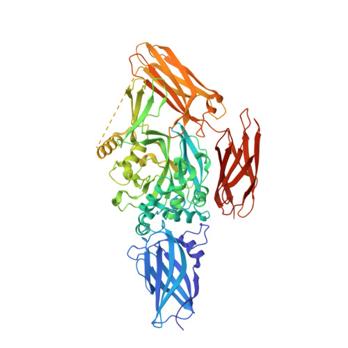
    1NUD, 1NUF, 1NUG

  • PubMed Abstract: 

    The transglutaminase 3 enzyme is widely expressed in many tissues including epithelia. We have shown previously that it can bind three Ca2+ ions, which in site one is constitutively bound, while those in sites two and three are acquired during activation and are required for activity. In particular, binding at site three opens a channel through the enzyme and exposes two tryptophan residues near the active site that are thought to be important for enzyme reaction. In this study, we have solved the structures of three more forms of this enzyme by x-ray crystallography in the presence of Ca2+ and/or Mg2+, which provide new insights on the precise contribution of each Ca2+ ion to activation and activity. First, we found that Ca2+ ion in site one can be exchanged with difficulty, and it has a binding affinity of Kd = 0.3 microm (DeltaH = -6.70 +/- 0.52 kcal/mol), which suggests it is important for the stabilization of the enzyme. Site two can be occupied by some lanthanides but only Ca2+ of the Group 2 family of alkali earth metals, and its occupancy are required for activity. Site three can be occupied by some lanthanides, Ca2+,or Mg2+; however, when Mg2+ is present, the enzyme is inactive, and the channel is closed. Thus Ca2+ binding in both sites two and three cooperate in opening the channel. We speculate that manipulation of the channel opening could be controlled by intracellular cation levels. Together, these data have important implications for reaction mechanism of the enzyme: the opening of a channel perhaps controls access to and manipulation of substrates at the active site.


  • Organizational Affiliation

    Laboratory of Skin Biology, the Laboratory of X-ray Crystallography/Office of Science and Technology, National Institute of Health, Bethesda, Maryland 20892-8023, USA. Bijan@discus.niams.nih.gov


Macromolecules
Find similar proteins by:  (by identity cutoff)  |  3D Structure
Entity ID: 1
MoleculeChains Sequence LengthOrganismDetailsImage
Protein-glutamine glutamyltransferase E
A, B
692Homo sapiensMutation(s): 1 
Gene Names: TGM3
EC: 2.3.2.13
UniProt & NIH Common Fund Data Resources
Find proteins for Q08188 (Homo sapiens)
Explore Q08188 
Go to UniProtKB:  Q08188
PHAROS:  Q08188
GTEx:  ENSG00000125780 
Entity Groups  
Sequence Clusters30% Identity50% Identity70% Identity90% Identity95% Identity100% Identity
UniProt GroupQ08188
Sequence Annotations
Expand
  • Reference Sequence
Small Molecules
Ligands 3 Unique
IDChains Name / Formula / InChI Key2D Diagram3D Interactions
BR
Query on BR

Download Ideal Coordinates CCD File 
J [auth A]
K [auth A]
L [auth A]
M [auth A]
U [auth B]
J [auth A],
K [auth A],
L [auth A],
M [auth A],
U [auth B],
V [auth B],
W [auth B],
X [auth B],
Y [auth B]
BROMIDE ION
Br
CPELXLSAUQHCOX-UHFFFAOYSA-M
CA
Query on CA

Download Ideal Coordinates CCD File 
C [auth A]
D [auth A]
E [auth A]
N [auth B]
O [auth B]
C [auth A],
D [auth A],
E [auth A],
N [auth B],
O [auth B],
P [auth B]
CALCIUM ION
Ca
BHPQYMZQTOCNFJ-UHFFFAOYSA-N
CL
Query on CL

Download Ideal Coordinates CCD File 
F [auth A]
G [auth A]
H [auth A]
I [auth A]
Q [auth B]
F [auth A],
G [auth A],
H [auth A],
I [auth A],
Q [auth B],
R [auth B],
S [auth B],
T [auth B]
CHLORIDE ION
Cl
VEXZGXHMUGYJMC-UHFFFAOYSA-M
Experimental Data & Validation

Experimental Data

  • Method: X-RAY DIFFRACTION
  • Resolution: 2.70 Å
  • R-Value Free: 0.265 
  • R-Value Work: 0.199 
  • Space Group: P 1 21 1
Unit Cell:
Length ( Å )Angle ( ˚ )
a = 57.54α = 90
b = 115.37β = 92.66
c = 121.39γ = 90
Software Package:
Software NamePurpose
HKL-2000data collection
SCALEPACKdata scaling
AMoREphasing
CNSrefinement
HKL-2000data reduction

Structure Validation

View Full Validation Report


Entry History 

Deposition Data

  • Released Date: 2003-04-22 
  • Deposition Author(s): Ahvazi, B.

Revision History  (Full details and data files)

  • Version 1.0: 2003-04-22
    Type: Initial release
  • Version 1.1: 2008-04-29
    Changes: Version format compliance
  • Version 1.2: 2011-07-13
    Changes: Version format compliance
  • Version 1.3: 2021-10-27
    Changes: Database references, Derived calculations
  • Version 1.4: 2023-08-16
    Changes: Data collection, Refinement description