


Crystal Structure of Bacillus subtilis YojF protein
Kim, Y., Korolev, O., Joachimiak, A.To be published.
Experimental Data Snapshot
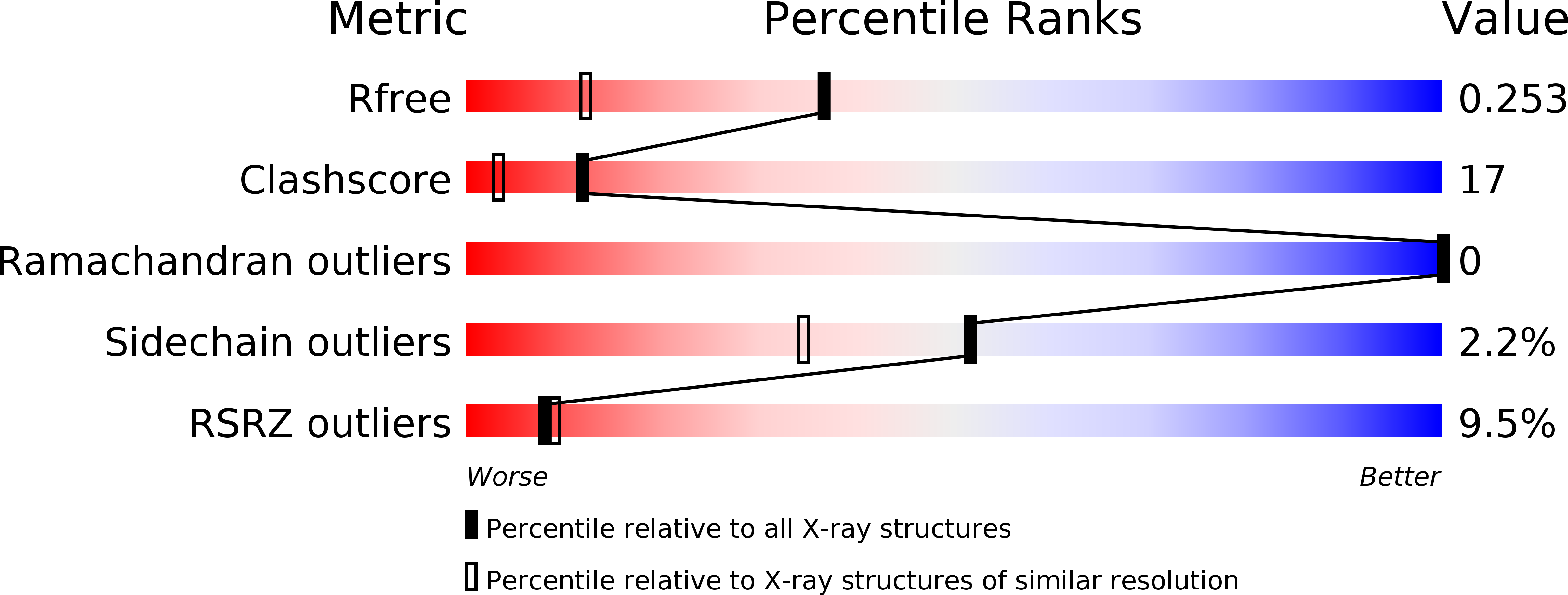
wwPDB Validation 3D Report Full Report
Entity ID: 1 | |||||
|---|---|---|---|---|---|
| Molecule | Chains | Sequence Length | Organism | Details | Image |
| protein yojF | 119 | Bacillus subtilis | Mutation(s): 3 Gene Names: YojF | 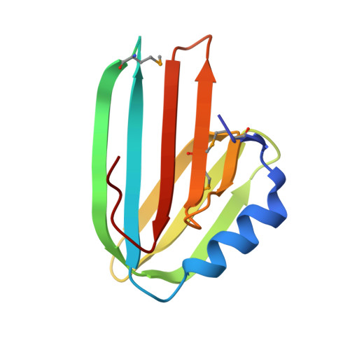 | |
UniProt | |||||
Find proteins for O31858 (Bacillus subtilis (strain 168)) Explore O31858 Go to UniProtKB: O31858 | |||||
Entity Groups | |||||
| Sequence Clusters | 30% Identity50% Identity70% Identity90% Identity95% Identity100% Identity | ||||
| UniProt Group | O31858 | ||||
Sequence AnnotationsExpand | |||||
| |||||
| Ligands 2 Unique | |||||
|---|---|---|---|---|---|
| ID | Chains | Name / Formula / InChI Key | 2D Diagram | 3D Interactions | |
| GOL Query on GOL | C [auth A] | GLYCEROL C3 H8 O3 PEDCQBHIVMGVHV-UHFFFAOYSA-N |  | ||
| IPA Query on IPA | B [auth A] | ISOPROPYL ALCOHOL C3 H8 O KFZMGEQAYNKOFK-UHFFFAOYSA-N |  | ||
| Modified Residues 1 Unique | |||||
|---|---|---|---|---|---|
| ID | Chains | Type | Formula | 2D Diagram | Parent |
| MSE Query on MSE | A | L-PEPTIDE LINKING | C5 H11 N O2 Se |  | MET |
| Length ( Å ) | Angle ( ˚ ) |
|---|---|
| a = 78.703 | α = 90 |
| b = 78.703 | β = 90 |
| c = 31.074 | γ = 120 |
| Software Name | Purpose |
|---|---|
| CNS | refinement |
| d*TREK | data reduction |
| HKL-2000 | data reduction |
| HKL-2000 | data scaling |
| SOLVE | phasing |
| RESOLVE | phasing |
RCSB PDB (citation) is hosted by
RCSB PDB is a member of the

