Dual Coenzyme Specificity of Photosynthetic Glyceraldehyde-3-phosphate Dehydrogenase Interpreted by the Crystal Structure of A(4) Isoform Complexed with NAD
Falini, G., Fermani, S., Ripamonti, A., Sabatino, P., Sparla, F., Pupillo, P., Trost, P.(2003) Biochemistry 42: 4631-4639
- PubMed: 12705826
- DOI: https://doi.org/10.1021/bi0272149
- Primary Citation of Related Structures:
1NBO - PubMed Abstract:
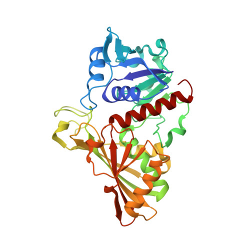
Photosynthetic glyceraldehyde-3-phosphate dehydrogenase (GAPDH) of Spinacia oleracea belongs to a wide group of GAPDHs found in most organisms displaying oxygenic photosynthesis, including cyanobacteria, green and red algae, and higher plants. As a major catalytic difference with respect to glycolytic GAPDH, photosynthetic GAPDH exhibits dual cofactor specificity toward pyridine nucleotides with a preference for NADP(H). Here we report the crystal structure of NAD-complexed recombinant A(4)-GAPDH (NAD-A(4)-GAPDH) from Spinacia oleracea, expressed in Escherichia coli. Its superimposition onto native A(4)-GAPDH complexed with NADP (NADP-A(4)-GAPDH) pinpoints specific conformational changes resulting from cofactor replacement. In photosynthetic NAD-A(4)-GAPDH, the side chain of Asp32 is oriented toward the coenzyme to interact with the adenine ribose diol, similar to glycolytic GAPDHs (NAD-specific). On the contrary, in NADP-A(4)-GAPDH Asp32 moves away to accommodate the additional 2'-phosphate group of the coenzyme and to minimize electrostatic repulsion. Asp32 rotation is allowed by the presence of the small residue Ala40, conserved in most photosynthetic GAPDHs, replacing bulky amino acid side chains in glycolytic GAPDHs. While in NADP-A(4)-GAPDH two amino acids, Thr33 and Ser188, are involved in hydrogen bonds with the 2'-phosphate group of NADP, in the NAD-complexed enzyme these interactions are lacking. The crystallographic structure of NAD-A(4)-GAPDH highlights that four residues, Thr33, Ala40, Ser188, and Ala187 (Leu, Leu, Pro, and Leu respectively, in glycolytic Bacillus stearothermophilus GAPDH sequence) are of primary importance for the dual cofactor specificity of photosynthetic GAPDH. These modifications seem to trace the minimum evolutionary route for a primitive NAD-specific GAPDH to be converted into the NADP-preferring enzyme of oxygenic photosynthetic organisms.
Organizational Affiliation:
Dipartimento di Chimica G. Ciamician, Alma Mater Studiorum Università di Bologna, 40126 Bologna, Italia.