1NAL

THE THREE-DIMENSIONAL STRUCTURE OF N-ACETYLNEURAMINATE LYASE FROM ESCHERICHIA COLI


Experimental Data Snapshot

  • Method: X-RAY DIFFRACTION
  • Resolution: 2.20 Å
  • R-Value Work: 0.208 
  • R-Value Observed: 0.208 

wwPDB Validation   3D Report Full Report


This is version 1.4 of the entry. See complete history


Literature

The three-dimensional structure of N-acetylneuraminate lyase from Escherichia coli.

Izard, T.Lawrence, M.C.Malby, R.L.Lilley, G.G.Colman, P.M.

(1994) Structure 2: 361-369

  • DOI: https://doi.org/10.1016/s0969-2126(00)00038-1
  • Primary Citation of Related Structures:  
    1NAL

  • PubMed Abstract: 

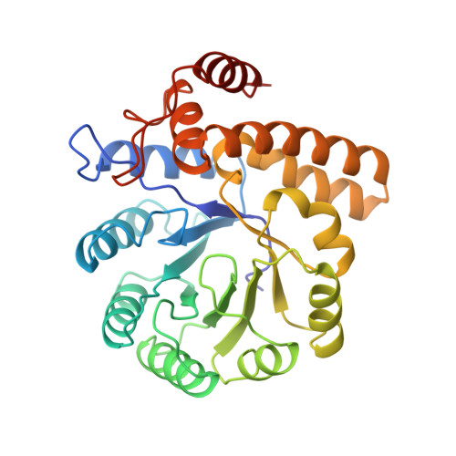
    N-acetylneuraminate lyase catalyzes the cleavage of N-acetylneuraminic acid (sialic acid) to form pyruvate and N-acetyl-D-mannosamine. The enzyme plays an important role in the regulation of sialic acid metabolism in bacteria. The reverse reaction can be exploited for the synthesis of sialic acid and some of its derivatives. The structure of the enzyme from Escherichia coli has been determined to 2.2 A resolution by X-ray crystallography. The enzyme is shown to be a tetramer, in which each subunit consists of an alpha/beta-barrel domain followed by a carboxy-terminal extension of three alpha-helices. The active site of the enzyme is tentatively identified as a pocket at the carboxy-terminal end of the eight-stranded beta-barrel. Lys165 lies within this pocket and is probably the reactive residue which forms a Schiff base intermediate with the substrate. The sequence of N-acetylneuraminate lyase has similarities to those of dihydrodipicolinate synthase and MosA (an enzyme implicated in rhizopine synthesis) suggesting that these last two enzymes share a similar structure to N-acetylneuraminate lyase.


  • Organizational Affiliation

    Biomolecular Research Institute, Parkville, Australia.


Macromolecules
Find similar proteins by:  (by identity cutoff)  |  3D Structure
Entity ID: 1
MoleculeChains Sequence LengthOrganismDetailsImage
N-ACETYLNEURAMINATE LYASEA [auth 1],
B [auth 2],
C [auth 3],
D [auth 4]
297Escherichia coliMutation(s): 0 
EC: 4.1.3.3
UniProt
Find proteins for P0A6L4 (Escherichia coli (strain K12))
Explore P0A6L4 
Go to UniProtKB:  P0A6L4
Entity Groups  
Sequence Clusters30% Identity50% Identity70% Identity90% Identity95% Identity100% Identity
UniProt GroupP0A6L4
Sequence Annotations
Expand
  • Reference Sequence
Experimental Data & Validation

Experimental Data

  • Method: X-RAY DIFFRACTION
  • Resolution: 2.20 Å
  • R-Value Work: 0.208 
  • R-Value Observed: 0.208 
  • Space Group: P 32 2 1
Unit Cell:
Length ( Å )Angle ( ˚ )
a = 122.8α = 90
b = 122.8β = 90
c = 198.8γ = 120
Software Package:
Software NamePurpose
X-PLORmodel building
X-PLORrefinement
X-PLORphasing

Structure Validation

View Full Validation Report


Entry History 

Deposition Data

Revision History  (Full details and data files)

  • Version 1.0: 1995-09-15
    Type: Initial release
  • Version 1.1: 2008-03-24
    Changes: Version format compliance
  • Version 1.2: 2011-07-13
    Changes: Version format compliance
  • Version 1.3: 2017-11-29
    Changes: Advisory, Derived calculations, Other
  • Version 1.4: 2024-02-14
    Changes: Advisory, Data collection, Database references, Derived calculations