A consensus structure for omega-conotoxins with different selectivities for voltage-sensitive calcium channel subtypes: comparison of MVIIA, SVIB and SNX-202.
Nielsen, K.J., Thomas, L., Lewis, R.J., Alewood, P.F., Craik, D.J.(1996) J Mol Biol 263: 297-310
- PubMed: 8913308
- DOI: https://doi.org/10.1006/jmbi.1996.0576
- Primary Citation of Related Structures:
1MVI, 1MVJ - PubMed Abstract:
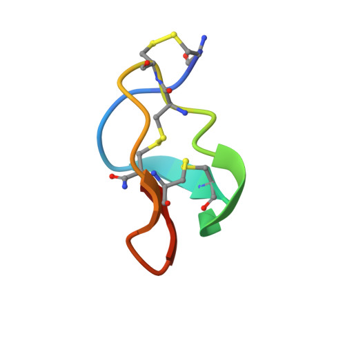
The omega-conotoxins are a set of structurally related peptides that have a wide range of specificities for different subtypes of the voltage-sensitive calcium channel (VSCC). To understand their VSCC subtype differentiation we studied the structure of two naturally occurring omega-conotoxins, MVIIA (specific to N-type) and SVIB (specific to P/Q-type) and a synthetic hybrid, SNX-202, which has altered specificities to both VSCC subtypes. The secondary structures of the three peptides are almost identical, consisting of a triple-stranded beta-sheet and several turns. A comparison of NMR data emphasizes the structural similarities between the peptides and highlights some minor structural differences. In the three-dimensional structures of SVIB and MVIIA these are manifested as orientational differences between two key loops. The structural rigidity of MVIIA was also examined. H alpha shifts are similar in a range of solvents, indicating that there are no solvent-induced changes in structure. The omega-conotoxins form a consensus structure despite differences in sequence and VSCC subtype specificity. This indicates that the omega-conotoxin macrosites for the N/P/Q-subfamily of VSCCs are related, with specificity for receptor targets being conferred by the positions of functional side-chains on the surface of the peptides.
Organizational Affiliation:
Centre for Drug Design and Development, University of Queensland, Brisbane, Australia.