Structural and kinetic studies of sugar binding to galactose mutarotase from Lactococcus lactis.
Thoden, J.B., Kim, J., Raushel, F.M., Holden, H.M.(2002) J Biol Chem 277: 45458-45465
- PubMed: 12218067
- DOI: https://doi.org/10.1074/jbc.M208395200
- Primary Citation of Related Structures:
1MMU, 1MMX, 1MMY, 1MMZ, 1MN0 - PubMed Abstract:
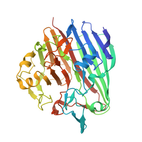
Galactose mutarotase catalyzes the conversion of beta-D-galactose to alpha-D-galactose in the Leloir pathway for galactose metabolism. The high resolution x-ray structure of the dimeric enzyme from Lactococcus lactis was recently solved and shown to be topologically similar to the 18-stranded, anti-parallel beta-motif observed for domain 5 of beta-galactosidase. In addition to determining the overall molecular fold of galactose mutarotase, this initial investigation also provided a detailed description of the electrostatic interactions between the enzyme and its physiologically relevant substrate, galactose. Specifically, the side chains of His-96 and His-170 were shown to be located within hydrogen bonding distance to the C-5 oxygen of the substrate, while the carboxylate of Glu-304 was positioned near the C-1 hydroxyl group of the sugar. On the basis of this initial study, a possible role for Glu-304 as the general acid/base group in catalysis was put forth. Here we describe the combined x-ray crystallographic and kinetic analyses of L. lactis galactose mutarotase complexed with D-glucose, D-fucose, D-quinovose, L-arabinose, or D-xylose. These investigations have revealed that there are several distinct binding modes for these sugars, which are dependent upon the spatial orientation of the C-4 hydroxyl group. In those sugars with the same C-4 hydroxyl group orientation as galactose, their C-1 hydroxyl groups are invariably located near Glu-304. For those sugars, which have the same C-4 hydroxyl group configuration as glucose, the C-1 hydroxyls are typically located near Asp-243. These different binding modes correlate with both the observed kinetic parameters and the presence or absence of a hydrogen bond between the guanidinium group of Arg-71 and the C-4 hydroxyl group of the sugar ligand.
Organizational Affiliation:
Department of Biochemistry, University of Wisconsin, Madison, Wisconsin 53706-1544, USA. JBThoden@facstaff.wisc.edu