1LGB

INTERACTION OF A LEGUME LECTIN WITH THE N2 FRAGMENT OF HUMAN LACTOTRANSFERRIN OR WITH THE ISOLATED BIANTENNARY GLYCOPEPTIDE: ROLE OF THE FUCOSE MOIETY


Experimental Data Snapshot

  • Method: X-RAY DIFFRACTION
  • Resolution: 3.30 Å
  • R-Value Work: 0.210 
  • R-Value Observed: 0.210 

wwPDB Validation   3D Report Full Report


This is version 2.0 of the entry. See complete history


Literature

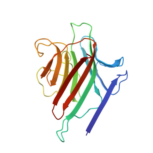
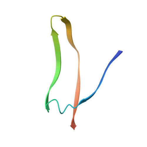
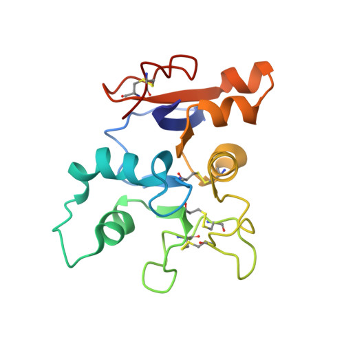
Structures of a legume lectin complexed with the human lactotransferrin N2 fragment, and with an isolated biantennary glycopeptide: role of the fucose moiety.

Bourne, Y.Mazurier, J.Legrand, D.Rouge, P.Montreuil, J.Spik, G.Cambillau, C.

(1994) Structure 2: 209-219

  • DOI: https://doi.org/10.1016/s0969-2126(00)00022-8
  • Primary Citation of Related Structures:  
    1LGB, 1LGC

  • PubMed Abstract: 

    Lectins mediate cell-cell interactions by specifically recognizing oligosaccharide chains. Legume lectins serve as mediators for the symbiotic interactions between plants and nitrogen-fixing microorganisms, an important process in the nitrogen cycle. Lectins from the Viciae tribe have a high affinity for the fucosylated biantennary N-acetyllactosamine-type glycans which are to be found in the majority of N-glycosylproteins. While the structures of several lectins complexed with incomplete oligosaccharides have been solved, no previous structure has included the complete glycoprotein. We have determined the crystal structures of Lathyrus ochrus isolectin II complexed with the N2 monoglycosylated fragment of human lactotransferrin (18 kDa) and an isolated glycopeptide (2.1 kDa) fragment of human lactotransferrin (at 3.3 A and 2.8 A resolution, respectively). Comparison between the two structures showed that the protein part of the glycoprotein has little influence on either the stabilization of the complex or the sugar conformation. In both cases the oligosaccharide adopts the same extended conformation. Besides the essential mannose moiety of the monosaccharide-binding site, the fucose-1' of the core has a large surface of interaction with the lectin. This oligosaccharide conformation differs substantially from that seen in the previously determined isolectin I-octasaccharide complex. Comparison of our structure with that of concanavalin A (ConA) suggests that the ConA binding site cannot accommodate this fucose. Our results explain the observation that Viciae lectins have a higher affinity for fucosylated oligosaccharides than for unfucosylated ones, whereas the affinity of ConA for these types of oligosaccharides is similar. This explanation is testable by mutagenesis experiments. Our structure shows a large complementary surface area between the oligosaccharide and the lectin, in contrast with the recently determined structure of a complex between the carbohydrate recognition domain of a C-type mammalian lectin and an oligomannoside, where only the non-reducing terminal mannose residue interacts with the lectin.


  • Organizational Affiliation

    Laboratoire de Cristallographie et de Cristallisation des Macromolécules Biologiques, CNRS URA 1296, Faculté de Médecine Secteur-Nord, Marseille, France.


Macromolecules
Find similar proteins by:  (by identity cutoff)  |  3D Structure
Entity ID: 1
MoleculeChains Sequence LengthOrganismDetailsImage
LEGUME ISOLECTIN II (ALPHA CHAIN)181Lathyrus ochrusMutation(s): 0 
UniProt
Find proteins for P04122 (Lathyrus ochrus)
Explore P04122 
Go to UniProtKB:  P04122
Entity Groups  
Sequence Clusters30% Identity50% Identity70% Identity90% Identity95% Identity100% Identity
UniProt GroupP04122
Sequence Annotations
Expand
  • Reference Sequence
Find similar proteins by:  (by identity cutoff)  |  3D Structure
Entity ID: 2
MoleculeChains Sequence LengthOrganismDetailsImage
LEGUME ISOLECTIN II (BETA CHAIN)53Lathyrus ochrusMutation(s): 0 
UniProt
Find proteins for P12307 (Lathyrus ochrus)
Explore P12307 
Go to UniProtKB:  P12307
Entity Groups  
Sequence Clusters30% Identity50% Identity70% Identity90% Identity95% Identity100% Identity
UniProt GroupP12307
Sequence Annotations
Expand
  • Reference Sequence
Find similar proteins by:  (by identity cutoff)  |  3D Structure
Entity ID: 3
MoleculeChains Sequence LengthOrganismDetailsImage
LACTOTRANSFERRIN (N2 FRAGMENT)159Homo sapiensMutation(s): 0 
UniProt & NIH Common Fund Data Resources
Find proteins for P02788 (Homo sapiens)
Explore P02788 
Go to UniProtKB:  P02788
PHAROS:  P02788
GTEx:  ENSG00000012223 
Entity Groups  
Sequence Clusters30% Identity50% Identity70% Identity90% Identity95% Identity100% Identity
UniProt GroupP02788
Sequence Annotations
Expand
  • Reference Sequence
Oligosaccharides

Help

Entity ID: 4
MoleculeChains Length2D Diagram Glycosylation3D Interactions
beta-D-galactopyranose-(1-4)-2-acetamido-2-deoxy-beta-D-glucopyranose-(1-2)-alpha-D-mannopyranose-(1-3)-[2-acetamido-2-deoxy-beta-D-glucopyranose-(1-2)-alpha-D-mannopyranose-(1-6)]beta-D-mannopyranose-(1-4)-2-acetamido-2-deoxy-beta-D-glucopyranose-(1-4)-[alpha-L-fucopyranose-(1-6)]2-acetamido-2-deoxy-beta-D-glucopyranose
D
9N-Glycosylation
Glycosylation Resources
GlyTouCan:  G58667NI
GlyCosmos:  G58667NI
GlyGen:  G58667NI
Small Molecules
Ligands 2 Unique
IDChains Name / Formula / InChI Key2D Diagram3D Interactions
MN
Query on MN

Download Ideal Coordinates CCD File 
E [auth A]MANGANESE (II) ION
Mn
WAEMQWOKJMHJLA-UHFFFAOYSA-N
CA
Query on CA

Download Ideal Coordinates CCD File 
F [auth A]CALCIUM ION
Ca
BHPQYMZQTOCNFJ-UHFFFAOYSA-N
Experimental Data & Validation

Experimental Data

  • Method: X-RAY DIFFRACTION
  • Resolution: 3.30 Å
  • R-Value Work: 0.210 
  • R-Value Observed: 0.210 
  • Space Group: P 43 21 2
Unit Cell:
Length ( Å )Angle ( ˚ )
a = 63.5α = 90
b = 63.5β = 90
c = 251.9γ = 90
Software Package:
Software NamePurpose
X-PLORmodel building
X-PLORrefinement
X-PLORphasing

Structure Validation

View Full Validation Report


Entry History 

Deposition Data

Revision History  (Full details and data files)

  • Version 1.0: 1994-08-31
    Type: Initial release
  • Version 1.1: 2008-03-03
    Changes: Version format compliance
  • Version 1.2: 2011-07-13
    Changes: Derived calculations, Version format compliance
  • Version 1.3: 2017-11-29
    Changes: Derived calculations, Other
  • Version 2.0: 2020-07-29
    Type: Remediation
    Reason: Carbohydrate remediation
    Changes: Atomic model, Data collection, Database references, Derived calculations, Structure summary