1K7C

Rhamnogalacturonan acetylesterase with seven N-linked carbohydrate residues distributed at two N-glycosylation sites refined at 1.12 A resolution


Experimental Data Snapshot

  • Method: X-RAY DIFFRACTION
  • Resolution: 1.12 Å
  • R-Value Free: 0.134 
  • R-Value Work: 0.103 
  • R-Value Observed: 0.104 

wwPDB Validation   3D Report Full Report


Ligand Structure Quality Assessment 


This is version 2.1 of the entry. See complete history


Literature

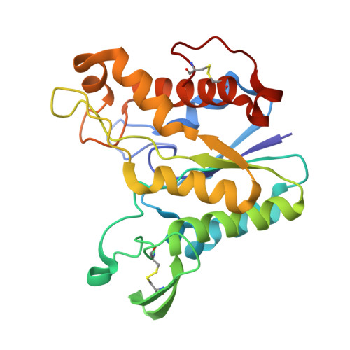
A branched N-linked glycan at atomic resolution in the 1.12 A structure of rhamnogalacturonan acetylesterase.

Molgaard, A.Larsen, S.

(2002) Acta Crystallogr D Biol Crystallogr 58: 111-119

  • DOI: https://doi.org/10.1107/s0907444901018479
  • Primary Citation of Related Structures:  
    1K7C

  • PubMed Abstract: 

    The crystal structure of the glycoprotein rhamnogalacturonan acetylesterase from Aspergillus aculeatus has been refined to a resolution of 1.12 A using synchrotron data collected at 263 K. Both of the two putative N-glycosylation sites at Asn104 and Asn182 are glycosylated and, owing to crystal contacts, the glycan structure at Asn182 is exceptionally well defined in the electron-density maps, showing the six-carbohydrate structure Manalpha1-6(Manalpha1-3)Manalpha1-6Manbeta1-4GlcNAcbeta1-4GlcNAcbeta-Asn182. Equivalent carbohydrate residues were restrained to have similar geometries, but were refined without target values. The refined bond lengths and angles were compared with the values obtained from small-molecule studies that form the basis for the dictionaries used for glycoprotein refinement.


  • Organizational Affiliation

    Centre for Crystallographic Studies, University of Copenhagen, Universitetsparken 5, DK-2100 Copenhagen, Denmark.


Macromolecules
Find similar proteins by:  (by identity cutoff)  |  3D Structure
Entity ID: 1
MoleculeChains Sequence LengthOrganismDetailsImage
rhamnogalacturonan acetylesterase233Aspergillus aculeatusMutation(s): 0 
UniProt
Find proteins for Q00017 (Aspergillus aculeatus)
Explore Q00017 
Go to UniProtKB:  Q00017
Entity Groups  
Sequence Clusters30% Identity50% Identity70% Identity90% Identity95% Identity100% Identity
UniProt GroupQ00017
Sequence Annotations
Expand
  • Reference Sequence
Oligosaccharides

Help

Entity ID: 2
MoleculeChains Length2D Diagram Glycosylation3D Interactions
alpha-D-mannopyranose-(1-3)-[alpha-D-mannopyranose-(1-6)]alpha-D-mannopyranose-(1-6)-alpha-D-mannopyranose-(1-4)-2-acetamido-2-deoxy-beta-D-glucopyranose-(1-4)-2-acetamido-2-deoxy-beta-D-glucopyranose
B
6N-Glycosylation
Glycosylation Resources
GlyTouCan:  G17987PO
GlyCosmos:  G17987PO
GlyGen:  G17987PO
Experimental Data & Validation

Experimental Data

  • Method: X-RAY DIFFRACTION
  • Resolution: 1.12 Å
  • R-Value Free: 0.134 
  • R-Value Work: 0.103 
  • R-Value Observed: 0.104 
  • Space Group: P 21 21 21
Unit Cell:
Length ( Å )Angle ( ˚ )
a = 52.17α = 90
b = 56.92β = 90
c = 71.69γ = 90
Software Package:
Software NamePurpose
MAR345data collection
SCALEPACKdata scaling
MLPHAREphasing
SHELXL-97refinement

Structure Validation

View Full Validation Report


Ligand Structure Quality Assessment 


Entry History 

Deposition Data

Revision History  (Full details and data files)

  • Version 1.0: 2001-12-28
    Type: Initial release
  • Version 1.1: 2008-04-27
    Changes: Version format compliance
  • Version 1.2: 2011-07-13
    Changes: Non-polymer description, Version format compliance
  • Version 2.0: 2020-07-29
    Type: Remediation
    Reason: Carbohydrate remediation
    Changes: Advisory, Atomic model, Data collection, Derived calculations, Refinement description, Structure summary
  • Version 2.1: 2023-10-25
    Changes: Data collection, Database references, Refinement description, Structure summary