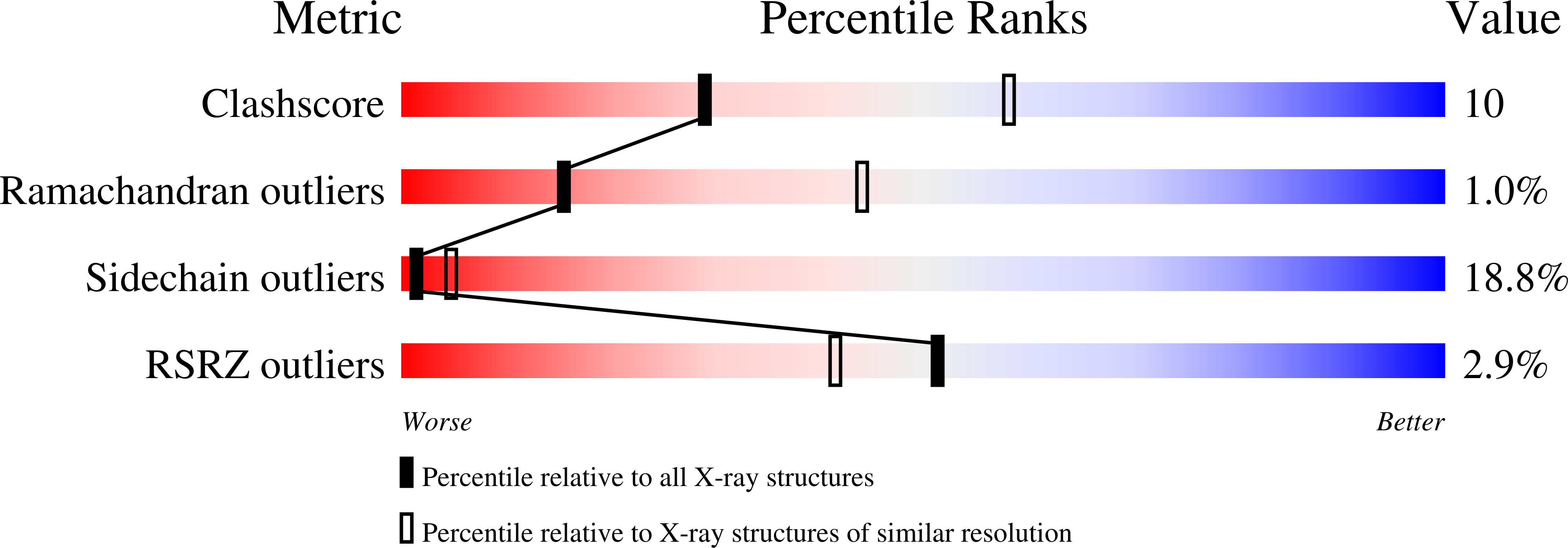
1JYM

Crystals of Peptide Deformylase from Plasmodium falciparum with Ten Subunits per Asymmetric Unit Reveal Critical Characteristics of the Active Site for Drug Design


Experimental Data Snapshot

  • Method: X-RAY DIFFRACTION
  • Resolution: 2.80 Å
  • R-Value Free: 0.296 
  • R-Value Work: 0.231 
  • R-Value Observed: 0.234 

wwPDB Validation   3D Report Full Report


This is version 1.4 of the entry. See complete history


Literature

Crystals of peptide deformylase from Plasmodium falciparum reveal critical characteristics of the active site for drug design.

Kumar, A.Nguyen, K.T.Srivathsan, S.Ornstein, B.Turley, S.Hirsh, I.Pei, D.Hol, W.G.

(2002) Structure 10: 357-367

  • DOI: https://doi.org/10.1016/s0969-2126(02)00719-0
  • Primary Citation of Related Structures:  
    1JYM

  • PubMed Abstract: 

    Peptide deformylase catalyzes the deformylation reaction of the amino terminal fMet residue of newly synthesized proteins in bacteria, and most likely in Plasmodium falciparum, and has therefore been identified as a potential antibacterial and antimalarial drug target. The structure of P. falciparum peptide deformylase, determined at 2.8 A resolution with ten subunits per asymmetric unit, is similar to the bacterial enzyme with the residues involved in catalysis, the position of the bound metal ion, and a catalytically important water structurally conserved between the two enzymes. However, critical differences in the substrate binding region explain the poor affinity of E. coli deformylase inhibitors and substrates toward the Plasmodium enzyme. The Plasmodium structure serves as a guide for designing novel antimalarials.


  • Organizational Affiliation

    Howard Hughes Medical Institute, University of Washington, Seattle 98195, USA.


Macromolecules
Find similar proteins by:  (by identity cutoff)  |  3D Structure
Entity ID: 1
MoleculeChains Sequence LengthOrganismDetailsImage
Peptide Deformylase
A, B, C, D, E
A, B, C, D, E, F, G, H, I, J
183Plasmodium falciparumMutation(s): 0 
Gene Names: PDF
EC: 3.5.1.31
UniProt
Find proteins for Q8I372 (Plasmodium falciparum (isolate 3D7))
Explore Q8I372 
Go to UniProtKB:  Q8I372
Entity Groups  
Sequence Clusters30% Identity50% Identity70% Identity90% Identity95% Identity100% Identity
UniProt GroupQ8I372
Sequence Annotations
Expand
  • Reference Sequence
Small Molecules
Modified Residues  1 Unique
IDChains TypeFormula2D DiagramParent
MSE
Query on MSE
A, B, C, D, E
A, B, C, D, E, F, G, H, I, J
L-PEPTIDE LINKINGC5 H11 N O2 SeMET
Experimental Data & Validation

Experimental Data

  • Method: X-RAY DIFFRACTION
  • Resolution: 2.80 Å
  • R-Value Free: 0.296 
  • R-Value Work: 0.231 
  • R-Value Observed: 0.234 
  • Space Group: P 41
Unit Cell:
Length ( Å )Angle ( ˚ )
a = 121.263α = 90
b = 121.263β = 90
c = 177.272γ = 90
Software Package:
Software NamePurpose
DENZOdata reduction
SCALEPACKdata scaling
SOLVEphasing
REFMACrefinement

Structure Validation

View Full Validation Report


Entry History 

Deposition Data

Revision History  (Full details and data files)

  • Version 1.0: 2002-03-13
    Type: Initial release
  • Version 1.1: 2008-05-05
    Changes: Version format compliance
  • Version 1.2: 2011-07-13
    Changes: Version format compliance
  • Version 1.3: 2023-08-16
    Changes: Data collection, Database references, Derived calculations, Refinement description
  • Version 1.4: 2023-11-15
    Changes: Data collection