1JHK

Crystal structure of the anti-estradiol antibody 57-2


Experimental Data Snapshot

  • Method: X-RAY DIFFRACTION
  • Resolution: 2.51 Å
  • R-Value Free: 0.262 
  • R-Value Work: 0.198 
  • R-Value Observed: 0.230 

wwPDB Validation   3D Report Full Report


This is version 1.3 of the entry. See complete history


Literature

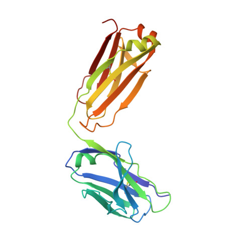
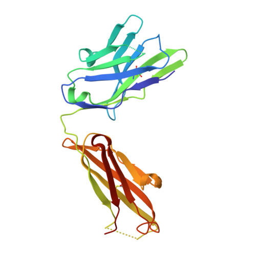
Crystal structure of a recombinant anti-estradiol Fab fragment in complex with 17beta -estradiol.

Lamminmaki, U.Kankare, J.A.

(2001) J Biol Chem 276: 36687-36694

  • DOI: https://doi.org/10.1074/jbc.M102367200
  • Primary Citation of Related Structures:  
    1JGL, 1JHK

  • PubMed Abstract: 

    The crystal structure of a Fab fragment of an anti-17beta-estradiol antibody 57-2 was determined in the absence and presence of the steroid ligand, 17beta-estradiol (E2), at 2.5 and 2.15-A resolutions, respectively. The antibody binds the steroid in a deep hydrophobic pocket formed at the interface between the variable domains. No major structural rearrangements take place upon ligand binding; however, a large part of the heavy chain variable domain near the binding pocket is unusually flexible and is partly stabilized when the steroid is bound. The nonpolar steroid skeleton of E2 is recognized by a number of hydrophobic interactions, whereas the two hydroxyl groups of E2 are hydrogen-bonded to the protein. Especially, the 17-hydroxyl group of E2 is recognized by an intricate hydrogen bonding network in which the 17-hydroxyl itself forms a rare four-center hydrogen bond with three polar amino acids; this hydrogen bonding arrangement accounts for the low cross-reactivity of the antibody with other estrogens such as estrone. The CDRH3 loop plays a prominent role in ligand binding. All the complementarity-determining regions of the light chain make direct contacts with the steroid, even CDRL2, which is rarely directly involved in the binding of haptens.


  • Organizational Affiliation

    Department of Biotechnology, University of Turku, Tykistökatu 6, 6th floor, 20520 Turku, Finland. urpo.lamminmaki@utu.fi


Macromolecules
Find similar proteins by:  (by identity cutoff)  |  3D Structure
Entity ID: 1
MoleculeChains Sequence LengthOrganismDetailsImage
Ig kappa-chainA [auth L]214Mus musculusMutation(s): 0 
UniProt
Find proteins for Q7TS98 (Mus musculus)
Explore Q7TS98 
Go to UniProtKB:  Q7TS98
Entity Groups  
Sequence Clusters30% Identity50% Identity70% Identity90% Identity95% Identity100% Identity
UniProt GroupQ7TS98
Sequence Annotations
Expand
  • Reference Sequence
Find similar proteins by:  (by identity cutoff)  |  3D Structure
Entity ID: 2
MoleculeChains Sequence LengthOrganismDetailsImage
Ig gamma-1-chainB [auth H]218Mus musculusMutation(s): 0 
UniProt
Find proteins for Q99LC4 (Mus musculus)
Explore Q99LC4 
Go to UniProtKB:  Q99LC4
Entity Groups  
Sequence Clusters30% Identity50% Identity70% Identity90% Identity95% Identity100% Identity
UniProt GroupQ99LC4
Sequence Annotations
Expand
  • Reference Sequence
Experimental Data & Validation

Experimental Data

  • Method: X-RAY DIFFRACTION
  • Resolution: 2.51 Å
  • R-Value Free: 0.262 
  • R-Value Work: 0.198 
  • R-Value Observed: 0.230 
  • Space Group: P 21 21 21
Unit Cell:
Length ( Å )Angle ( ˚ )
a = 48.267α = 90
b = 64.456β = 90
c = 165.702γ = 90
Software Package:
Software NamePurpose
DENZOdata reduction
SCALEPACKdata scaling
CNSrefinement
CNSphasing

Structure Validation

View Full Validation Report


Entry History 

Deposition Data

Revision History  (Full details and data files)

  • Version 1.0: 2001-10-10
    Type: Initial release
  • Version 1.1: 2008-04-27
    Changes: Version format compliance
  • Version 1.2: 2011-07-13
    Changes: Version format compliance
  • Version 1.3: 2023-10-25
    Changes: Data collection, Database references, Refinement description