1JEO

Crystal Structure of the Hypothetical Protein MJ1247 from Methanococcus jannaschii at 2.0 A Resolution Infers a Molecular Function of 3-Hexulose-6-Phosphate isomerase.


Experimental Data Snapshot

  • Method: X-RAY DIFFRACTION
  • Resolution: 2.00 Å
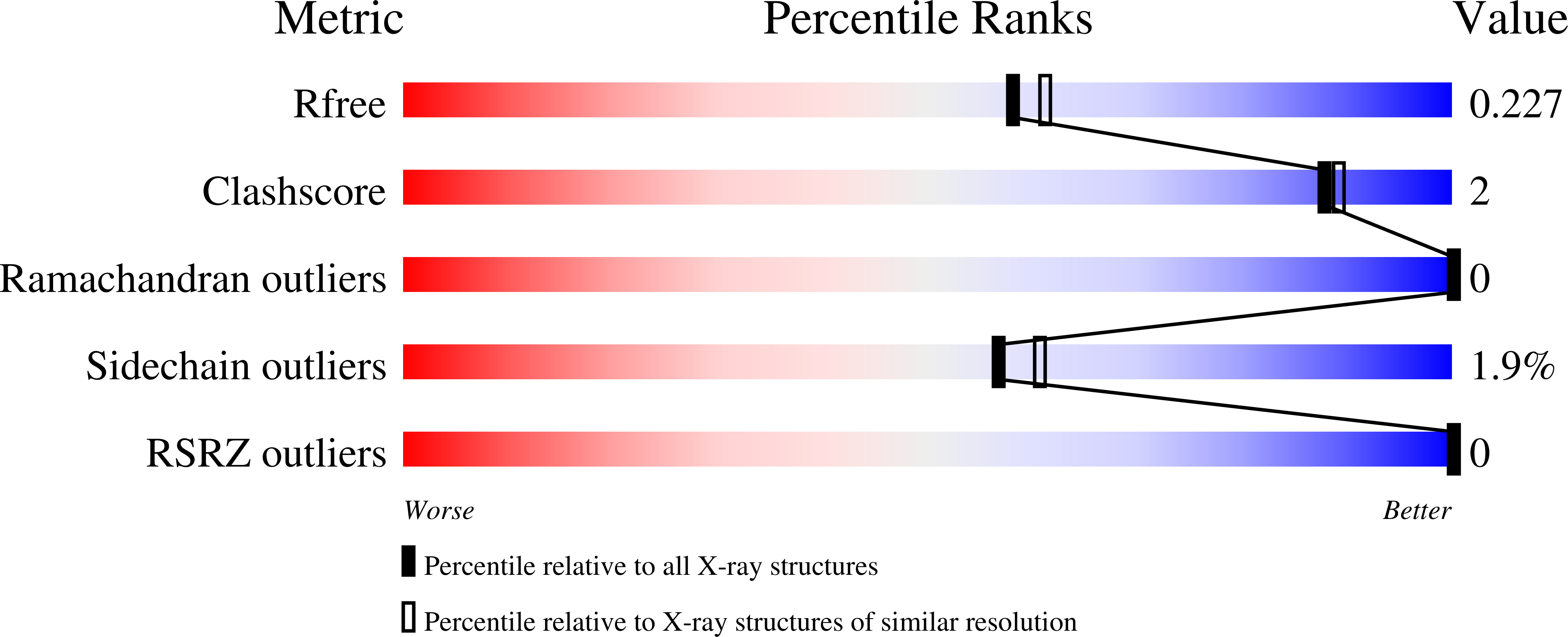
  • R-Value Free: 0.227 
  • R-Value Work: 0.189 
  • R-Value Observed: 0.190 

wwPDB Validation   3D Report Full Report


This is version 1.3 of the entry. See complete history


Literature

Crystal structure of MJ1247 protein from M. jannaschii at 2.0 A resolution infers a molecular function of 3-hexulose-6-phosphate isomerase.

Martinez-Cruz, L.A.Dreyer, M.K.Boisvert, D.C.Yokota, H.Martinez-Chantar, M.L.Kim, R.Kim, S.H.

(2002) Structure 10: 195-204

  • DOI: https://doi.org/10.1016/s0969-2126(02)00701-3
  • Primary Citation of Related Structures:  
    1JEO

  • PubMed Abstract: 

    The crystal structure of the hypothetical protein MJ1247 from Methanococccus jannaschii at 2 A resolution, a detailed sequence analysis, and biochemical assays infer its molecular function to be 3-hexulose-6-phosphate isomerase (PHI). In the dissimilatory ribulose monophosphate (RuMP) cycle, ribulose-5-phosphate is coupled to formaldehyde by the 3-hexulose-6-phosphate synthase (HPS), yielding hexulose-6-phosphate, which is then isomerized to fructose-6-phosphate by the enzyme 3-hexulose-6-phosphate isomerase. MJ1247 is an alpha/beta structure consisting of a five-stranded parallel beta sheet flanked on both sides by alpha helices, forming a three-layered alpha-beta-alpha sandwich. The fold represents the nucleotide binding motif of a flavodoxin type. MJ1247 is a tetramer in the crystal and in solution and each monomer has a folding similar to the isomerase domain of glucosamine-6-phosphate synthase (GlmS).


  • Organizational Affiliation

    Physical Biosciences Division, Lawrence Berkeley National Laboratory and Department of Chemistry, University of California, Berkeley, 94720, USA. almartinez@unav.es


Macromolecules
Find similar proteins by:  (by identity cutoff)  |  3D Structure
Entity ID: 1
MoleculeChains Sequence LengthOrganismDetailsImage
HYPOTHETICAL PROTEIN MJ1247180Methanocaldococcus jannaschiiMutation(s): 3 
Gene Names: mj1247
UniProt
Find proteins for Q58644 (Methanocaldococcus jannaschii (strain ATCC 43067 / DSM 2661 / JAL-1 / JCM 10045 / NBRC 100440))
Explore Q58644 
Go to UniProtKB:  Q58644
Entity Groups  
Sequence Clusters30% Identity50% Identity70% Identity90% Identity95% Identity100% Identity
UniProt GroupQ58644
Sequence Annotations
Expand
  • Reference Sequence
Small Molecules
Ligands 1 Unique
IDChains Name / Formula / InChI Key2D Diagram3D Interactions
CIT
Query on CIT

Download Ideal Coordinates CCD File 
B [auth A]CITRIC ACID
C6 H8 O7
KRKNYBCHXYNGOX-UHFFFAOYSA-N
Modified Residues  1 Unique
IDChains TypeFormula2D DiagramParent
CME
Query on CME
A
L-PEPTIDE LINKINGC5 H11 N O3 S2CYS
Experimental Data & Validation

Experimental Data

  • Method: X-RAY DIFFRACTION
  • Resolution: 2.00 Å
  • R-Value Free: 0.227 
  • R-Value Work: 0.189 
  • R-Value Observed: 0.190 
  • Space Group: I 2 2 2
Unit Cell:
Length ( Å )Angle ( ˚ )
a = 67.921α = 90
b = 68.155β = 90
c = 83.336γ = 90
Software Package:
Software NamePurpose
EPMRphasing
CNSrefinement
DENZOdata reduction
SCALEPACKdata scaling

Structure Validation

View Full Validation Report


Entry History 

Revision History  (Full details and data files)

  • Version 1.0: 2002-02-20
    Type: Initial release
  • Version 1.1: 2008-04-27
    Changes: Version format compliance
  • Version 1.2: 2011-07-13
    Changes: Derived calculations, Version format compliance
  • Version 1.3: 2024-04-03
    Changes: Data collection, Database references, Derived calculations, Refinement description