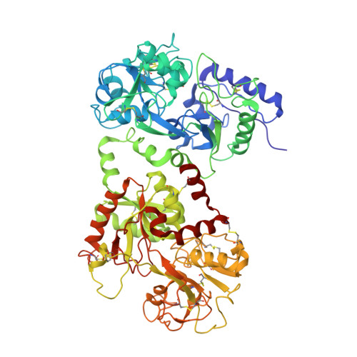
Crystal structure of equine apolactoferrin at 303 K providing further evidence of closed conformations of N and C lobes.
Kumar, P., Khan, J.A., Yadav, S., Singh, T.P.(2002) Acta Crystallogr D Biol Crystallogr 58: 225-232
- PubMed: 11807246
- DOI: https://doi.org/10.1107/s090744490101993x
- Primary Citation of Related Structures:
1I6B - PubMed Abstract:
Lactoferrin is an iron-binding protein. In the iron-bound state, the two domains of each lobe are invariably closed over an Fe(3+) ion. On the other hand, the structures of iron-free forms of lactoferrins from various species show different domain orientations. In order to determine the effects of external conditions such as pH, temperature and the presence of other additive agents on the crystal packing and consequently the influence of crystal packing forces on the final conformations of the two lobes of apolactoferrin, the structure of equine apolactoferrin has been determined at 303 K. The equine apolactoferrin was crystallized at 303 K using a microdialysis setup in which the concentration of protein was kept at 70 mg ml(-1) in 0.025 M Tris-HCl pH 8.0 with a reservoir containing 19% ethanol in the same buffer. The structure has been determined by molecular replacement using equine diferric lactoferrin as a model and was refined to an R factor of 0.23. The value of the overall B factor in the present structure is 81.3 A(2). The overall structure of the protein is similar to its earlier structure based on crystals grown at 277 K as well as that of diferric equine lactoferrin. The N and C lobes have been found to be slightly differently oriented (4.9 and 7.1 degrees, respectively) compared with the structures of equine diferric lactoferrin and apolactoferrin analyzed at 277 K, but the domain orientations in the two structures are identical as they remain closed over the empty iron-binding cleft. Overall, the structures of equine diferric lactoferrin, equine apolactoferrin at 277 K and the present structure of equine apolactoferrin at 303 K display identical domain orientations, suggesting that variation in the temperature of crystal growth, data collection and the processes of iron binding and iron release do not influence the arrangements of domains in equine lactoferrin.
Organizational Affiliation:
Department of Biophysics, All India Institute of Medical Sciences, New Delhi 110 029, India.