1GBG

BACILLUS LICHENIFORMIS BETA-GLUCANASE


Experimental Data Snapshot

  • Method: X-RAY DIFFRACTION
  • Resolution: 1.80 Å
  • R-Value Free: 0.236 
  • R-Value Observed: 0.165 

wwPDB Validation   3D Report Full Report


This is version 1.2 of the entry. See complete history


Literature

Crystal structure of Bacillus licheniformis 1,3-1,4-beta-D-glucan 4-glucanohydrolase at 1.8 A resolution.

Hahn, M.Pons, J.Planas, A.Querol, E.Heinemann, U.

(1995) FEBS Lett 374: 221-224

  • DOI: https://doi.org/10.1016/0014-5793(95)01111-q
  • Primary Citation of Related Structures:  
    1GBG

  • PubMed Abstract: 

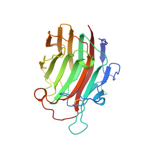
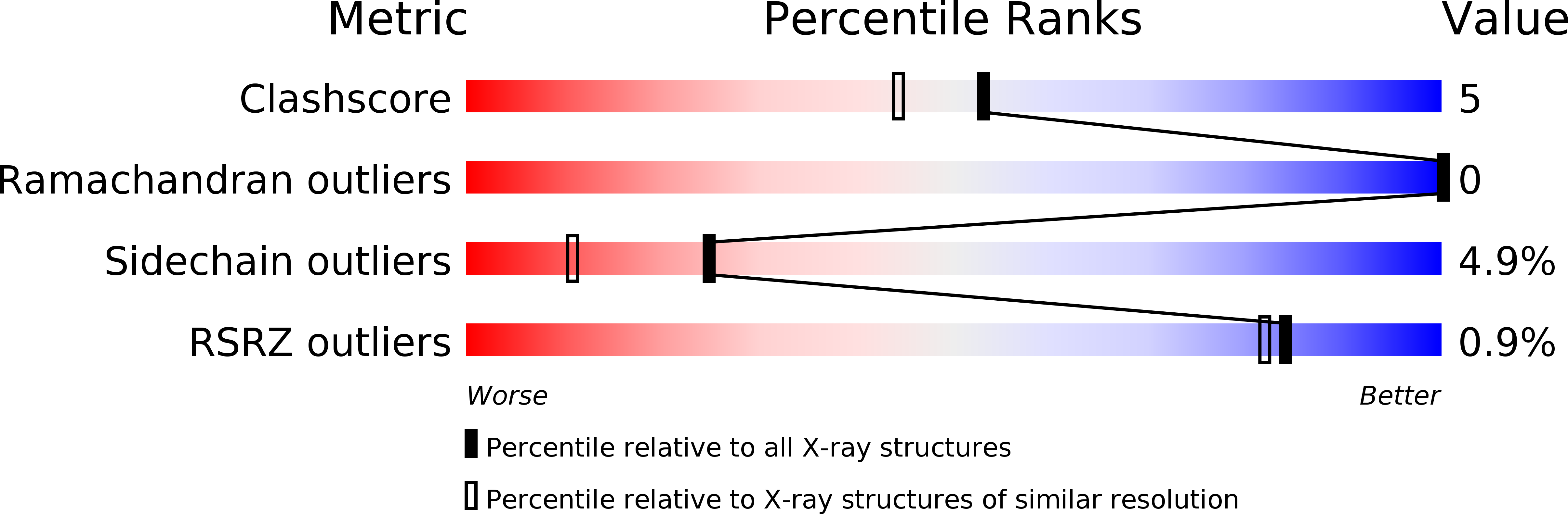
    The crystal structure of the 1,3-1,4-beta-D-glucan 4-glucanohydrolase from Bacillus licheniformis is solved at a resolution of 1.8 A and refined to R = 16.5%. The protein has a similar beta-sandwich structure as the homologous enzyme from Bacillus macerans and the hybrid H(A16-M). This demonstrates that the jellyroll fold of these proteins is remarkably rigid and only weakly influenced by crystal contacts. The crystal structure permits to extend mechanistic considerations derived for the B. licheniformis enzyme to the entire class of bacterial 1,3-1,4-beta-D-glucan 4-glucanohydrolases.


  • Organizational Affiliation

    Forschungsgruppe Kristallographie, Max-Delbrück-Centrum für Molekulare Medizin, Berlin, Germany.


Macromolecules
Find similar proteins by:  (by identity cutoff)  |  3D Structure
Entity ID: 1
MoleculeChains Sequence LengthOrganismDetailsImage
(1,3-1,4)-BETA-D-GLUCAN 4 GLUCANOHYDROLASE214Bacillus licheniformisMutation(s): 0 
EC: 3.2.1.73
UniProt
Find proteins for P27051 (Bacillus licheniformis)
Explore P27051 
Go to UniProtKB:  P27051
Entity Groups  
Sequence Clusters30% Identity50% Identity70% Identity90% Identity95% Identity100% Identity
UniProt GroupP27051
Sequence Annotations
Expand
  • Reference Sequence
Small Molecules
Ligands 1 Unique
IDChains Name / Formula / InChI Key2D Diagram3D Interactions
CA
Query on CA

Download Ideal Coordinates CCD File 
B [auth A]CALCIUM ION
Ca
BHPQYMZQTOCNFJ-UHFFFAOYSA-N
Experimental Data & Validation

Experimental Data

  • Method: X-RAY DIFFRACTION
  • Resolution: 1.80 Å
  • R-Value Free: 0.236 
  • R-Value Observed: 0.165 
  • Space Group: P 1
Unit Cell:
Length ( Å )Angle ( ˚ )
a = 35.33α = 64.66
b = 39.13β = 105.86
c = 43.88γ = 110.68
Software Package:
Software NamePurpose
MOSFLM/MADNESdata collection
TNTrefinement
MOSFLMdata reduction
MADNESdata reduction

Structure Validation

View Full Validation Report


Entry History 

Deposition Data

Revision History  (Full details and data files)

  • Version 1.0: 1995-12-07
    Type: Initial release
  • Version 1.1: 2008-03-03
    Changes: Version format compliance
  • Version 1.2: 2011-07-13
    Changes: Version format compliance