1G4B

CRYSTAL STRUCTURES OF THE HSLVU PEPTIDASE-ATPASE COMPLEX REVEAL AN ATP-DEPENDENT PROTEOLYSIS MECHANISM


Experimental Data Snapshot

  • Method: X-RAY DIFFRACTION
  • Resolution: 7.00 Å
  • R-Value Free: 0.432 
  • R-Value Work: 0.401 

wwPDB Validation   3D Report Full Report


This is version 1.3 of the entry. See complete history


Literature

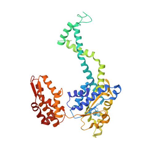
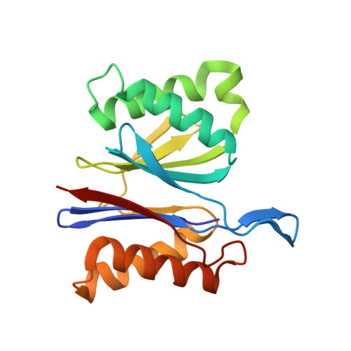
Crystal structures of the HslVU peptidase-ATPase complex reveal an ATP-dependent proteolysis mechanism.

Wang, J.Song, J.J.Franklin, M.C.Kamtekar, S.Im, Y.J.Rho, S.H.Seong, I.S.Lee, C.S.Chung, C.H.Eom, S.H.

(2001) Structure 9: 177-184

  • DOI: https://doi.org/10.1016/s0969-2126(01)00570-6
  • Primary Citation of Related Structures:  
    1G4A, 1G4B

  • PubMed Abstract: 

    The bacterial heat shock locus HslU ATPase and HslV peptidase together form an ATP-dependent HslVU protease. Bacterial HslVU is a homolog of the eukaryotic 26S proteasome. Crystallographic studies of HslVU should provide an understanding of ATP-dependent protein unfolding, translocation, and proteolysis by this and other ATP-dependent proteases. We present a 3.0 A resolution crystal structure of HslVU with an HslU hexamer bound at one end of an HslV dodecamer. The structure shows that the central pores of the ATPase and peptidase are next to each other and aligned. The central pore of HslU consists of a GYVG motif, which is conserved among protease-associated ATPases. The binding of one HslU hexamer to one end of an HslV dodecamer in the 3.0 A resolution structure opens both HslV central pores and induces asymmetric changes in HslV. Analysis of nucleotide binding induced conformational changes in the current and previous HslU structures suggests a protein unfolding-coupled translocation mechanism. In this mechanism, unfolded polypeptides are threaded through the aligned pores of the ATPase and peptidase and translocated into the peptidase central chamber.


  • Organizational Affiliation

    Department of Molecular Biophysics, Biochemistry, 266 Whitney Avenue, Yale University, 06520, New Haven, CT, USA. wang@mail.csb.yale.edu


Macromolecules
Find similar proteins by:  (by identity cutoff)  |  3D Structure
Entity ID: 1
MoleculeChains Sequence LengthOrganismDetailsImage
ATP-DEPENDENT HSL PROTEASE ATP-BINDING SUBUNIT HSLUA [auth E],
B [auth F],
C [auth K],
D [auth L]
443Escherichia coliMutation(s): 0 
UniProt
Find proteins for P0A6H5 (Escherichia coli (strain K12))
Explore P0A6H5 
Go to UniProtKB:  P0A6H5
Entity Groups  
Sequence Clusters30% Identity50% Identity70% Identity90% Identity95% Identity100% Identity
UniProt GroupP0A6H5
Sequence Annotations
Expand
  • Reference Sequence
Find similar proteins by:  (by identity cutoff)  |  3D Structure
Entity ID: 2
MoleculeChains Sequence LengthOrganismDetailsImage
ATP-DEPENDENT PROTEASE HSLVE [auth M],
F [auth N],
G [auth O],
H [auth P]
175Escherichia coliMutation(s): 0 
EC: 3.4.99
UniProt
Find proteins for P0A7B8 (Escherichia coli (strain K12))
Explore P0A7B8 
Go to UniProtKB:  P0A7B8
Entity Groups  
Sequence Clusters30% Identity50% Identity70% Identity90% Identity95% Identity100% Identity
UniProt GroupP0A7B8
Sequence Annotations
Expand
  • Reference Sequence
Experimental Data & Validation

Experimental Data

  • Method: X-RAY DIFFRACTION
  • Resolution: 7.00 Å
  • R-Value Free: 0.432 
  • R-Value Work: 0.401 
  • Space Group: P 3 2 1
Unit Cell:
Length ( Å )Angle ( ˚ )
a = 173.376α = 90
b = 173.376β = 90
c = 254.411γ = 120
Software Package:
Software NamePurpose
X-PLORmodel building
CNSrefinement
DENZOdata reduction
SCALEPACKdata scaling
X-PLORphasing

Structure Validation

View Full Validation Report


Entry History 

Deposition Data

Revision History  (Full details and data files)

  • Version 1.0: 2001-02-21
    Type: Initial release
  • Version 1.1: 2008-04-27
    Changes: Version format compliance
  • Version 1.2: 2011-07-13
    Changes: Version format compliance
  • Version 1.3: 2024-02-07
    Changes: Data collection, Database references